. 2019 Oct 15:13:1096.
doi: 10.3389/fnins.2019.01096. eCollection 2019.

Systemic and Intravitreal Antagonism of the TNFR1 Signaling Pathway Delays Axotomy-Induced Retinal Ganglion Cell Loss

Affiliations

Systemic and Intravitreal Antagonism of the TNFR1 Signaling Pathway Delays Axotomy-Induced Retinal Ganglion Cell Loss

Fernando Lucas-Ruiz et al. Front Neurosci. .

Abstract

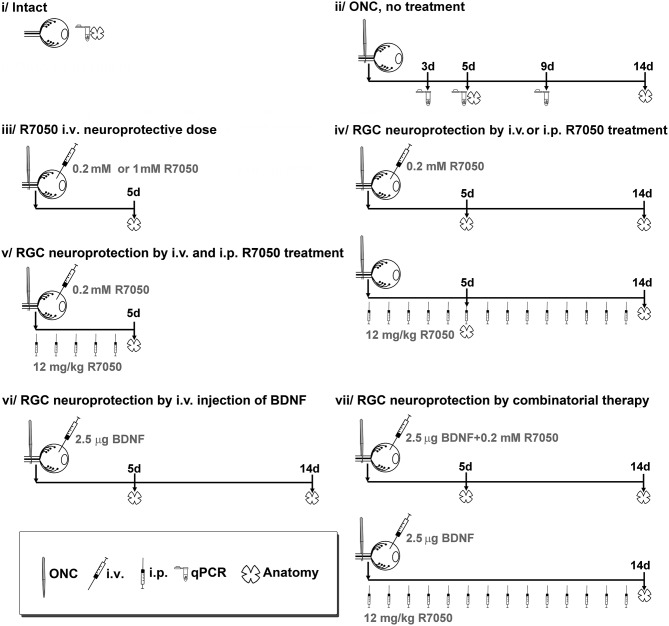
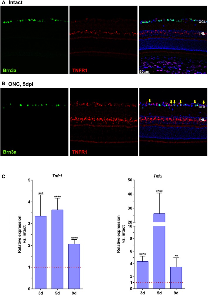
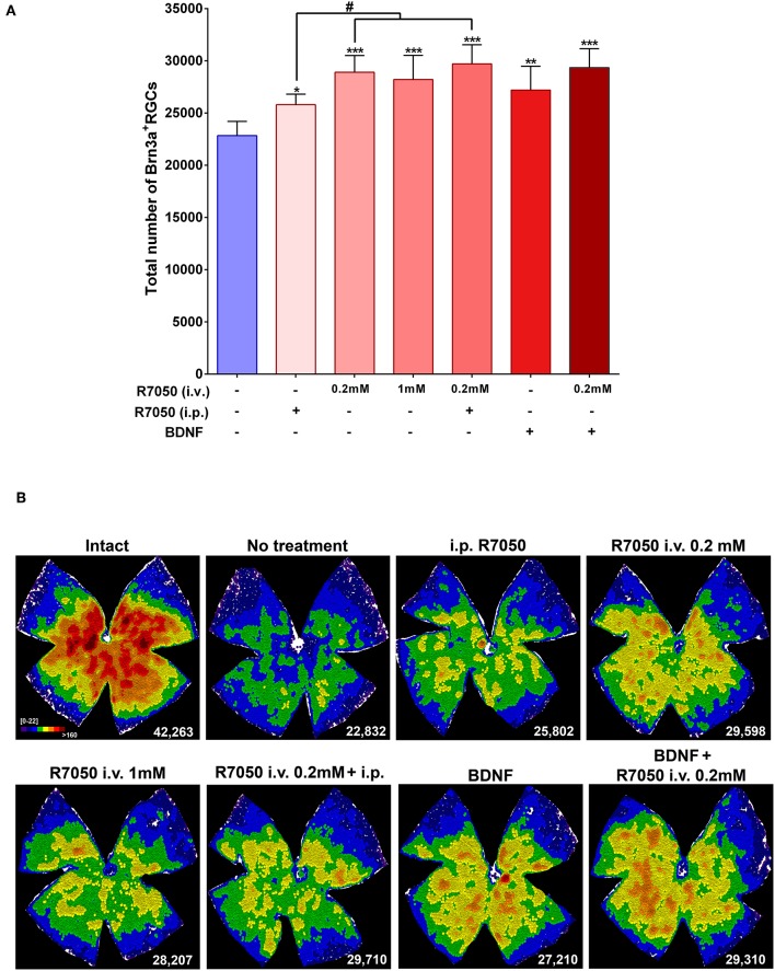
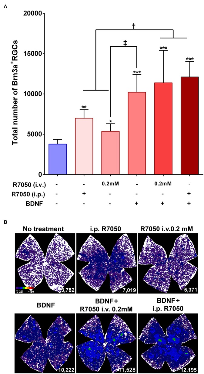
Here, we have blocked the signaling pathway of tumor necrosis factor α (TNFα) in a mouse model of traumatic neuropathy using a small cell permeable molecule (R7050) that inhibits TNFα/TNF receptor 1 (TNFR1) complex internalization. Adult pigmented mice were subjected to intraorbital optic nerve crush (ONC). Animals received daily intraperitoneal injections of R7050, and/or a single intravitreal administration the day of the surgery. Some animals received a combinatorial treatment with R7050 (systemic or local) and a single intravitreal injection of brain derived neurotrophic factor (BDNF). As controls, untreated animals were used. Retinas were analyzed for RGC survival 5 and 14 days after the lesion i.e., during the quick and slow phase of axotomy-induced RGC death. qPCR analyses were done to verify that Tnfr1 and TNFα were up-regulated after ONC. At 5 days post-lesion, R7050 intravitreal or systemic treatment neuroprotected RGCs as much as BDNF alone. At 14 days, RGC rescue by systemic or intravitreal administration of R7050 was similar. At this time point, intravitreal treatment with BDNF was significantly better than intravitreal R7050. Combinatory treatment was not better than BDNF alone, although at both time points, the mean number of surviving RGCs was higher. In conclusion, antagonism of the extrinsic pathway of apoptosis rescues axotomized RGCs as it does the activation of survival pathways by BDNF. However, manipulation of both pathways at the same time, does not improve RGC survival.

Keywords: BDNF; R7050; combinatory therapy; neuroprotection; optic nerve crush; retinal ganglion cells.


Figures

Figure 1
Figure 1
Experimental design and animal groups. R7050 toxicity was studied at 0.2 mM in intact retinas, which were analyzed 5 days after administration. As vehicle control groups, we assayed R7050 1 mM vehicle (5% DMSO in saline) since it has been published that at this concentration is toxic for neurons (Galvao et al., 2014). Two groups were done, intact + i.v. of 5% DMSO and ONC + i.v. of DMSO analyzed 5 days later. To reduce the number of animals, control groups for 1% DMSO (R7050 0.2 mM vehicle) were not done since in our conditions neither the population of RGCs nor their loss after axotomy was affected by an i.v. injection of 5% DMSO. Likewise, intraperitoneal vehicle of R7050 and intravitreal vehicle of BDNF groups were not done because we have already shown that those vehicle formulations and administration routes do not have a negative effect in the retina (Galindo-Romero et al., ; Di Pierdomenico et al., 2018). i.v, intravitreal injection; i.p., intraperitoneal injection.
Figure 2
Figure 2
TNFR1 is over-expressed in the retina after ONC. (A,B) Magnifications from retinal cross-sections showing the double immunodetection of Brn3a (green), TNFR1 (red), and the merged image with DAPI. In intact retinas (A) TNFR1 expression is observed mainly in the inner nuclear layer (INL), and in few Brn3a negative cells in the ganglion cell layer (GCL). Five days after ONC (B) many cells in the GCL, including RGCs (yellow arrows) express TNFR1. (C) Graph bars showing the fold change ± SEM of Tnfr1 and TNFα mRNA levels in ONC-injured retinas relative to intact retinas (value 1, red dotted line; **p < 0.01; ***p < 0.001; ****p < 0.0001, T-test vs. naive). d, days post-lesion.
Figure 3
Figure 3
Systemic and intravitreal treatment with R7050 alone or in combination with BDNF rescues RGCs during the quick phase of death after optic nerve axotomy. (A) Column bar graph showing the total mean number ± standard deviation of RGCs quantified in the different animal groups analyzed 5 days after ONC. *Statistically different from ONC alone (*p < 0.05; **p < 0.01; ***p < 0.001. ANOVA, Kruskal-Wallis. Dunn's post-hoc test). #p < 0.01 (T-test). For detailed statistics see Table 1. (B) Neighbor maps showing the distribution of RGCs in a representative retina from each group and treatment. These maps illustrate the number of RGCs around a given RGC in a radius of 0.200 mm with a color scale (top left panel) from 0 to 22 neighbors (purple) to >160 neighbors (dark red). At the bottom left of each map is shown the number of RGCs counted in the original retina.
Figure 4
Figure 4
Systemic and intravitreal treatment with R7050 alone or in combination with BDNF rescues RGCs during the slow phase of death after optic nerve axotomy. (A) Column bar graph showing the total mean number ± standard deviation of RGCs quantified in the different animal groups analyzed 14 days after ONC. *Statistically different from ONC alone (*p < 0.05; **p < 0.01; ***p < 0.001). p < 0.01; p < 0.05 (ANOVA, Kruskal-Wallis. Dunn's post-hoc test). See Table 1 for more details. (B) Neighbor maps showing the distribution of RGCs in a representative retina from each group and treatment. These maps illustrate the number of RGCs around a given RGC in a radius of 0.220 mm with a color scale (top left panel) from 0 to 22 neighbors (purple) to >160 neighbors (dark red). At the bottom left of each map is shown the number of RGCs counted in the original retina.

References

    1. Agudo M., Perez-Marin M. C., Lonngren U., Sobrado P., Conesa A., Canovas I., et al. . (2008). Time course profiling of the retinal transcriptome after optic nerve transection and optic nerve crush. Mol. Vis. 14, 1050–1063. - PMC - PubMed
    1. Agudo M., Perez-Marin M. C., Sobrado-Calvo P., Lonngren U., Salinas-Navarro M., Canovas I., et al. . (2009). Immediate upregulation of proteins belonging to different branches of the apoptotic cascade in the retina after optic nerve transection and optic nerve crush. Invest. Ophthalmol. Vis. Sci. 50, 424–431. 10.1167/iovs.08-2404 - DOI - PubMed
    1. Agudo-Barriuso M., Lahoz A., Nadal-Nicolas F. M., Sobrado-Calvo P., Piquer-Gil M., Diaz-Llopis M., et al. . (2013). Metabolomic changes in the rat retina after optic nerve crush. Invest. Ophthalmol. Vis. Sci. 54, 4249–4259. 10.1167/iovs.12-11451 - DOI - PubMed
    1. Cabal-Hierro L., Lazo P. S. (2012). Signal transduction by tumor necrosis factor receptors. Cell Signal. 24, 1297–1305. 10.1016/j.cellsig.2012.02.006 - DOI - PubMed
    1. Cheng X., Yang L., He P., Li R., Shen Y. (2010). Differential activation of tumor necrosis factor receptors distinguishes between brains from Alzheimer's disease and non-demented patients. J. Alzheimers Dis. 19, 621–630. 10.3233/JAD-2010-1253 - DOI - PMC - PubMed