. 2019 Oct 11:13:65.
doi: 10.3389/fncir.2019.00065. eCollection 2019.

The Organization of the Second Optic Chiasm of the Drosophila Optic Lobe

Affiliations

The Organization of the Second Optic Chiasm of the Drosophila Optic Lobe

Kazunori Shinomiya et al. Front Neural Circuits. .

Abstract

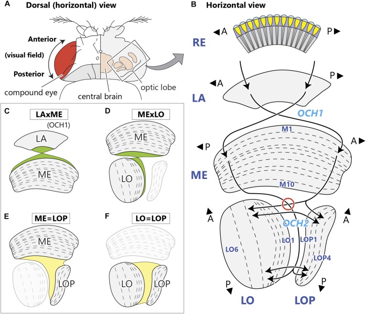
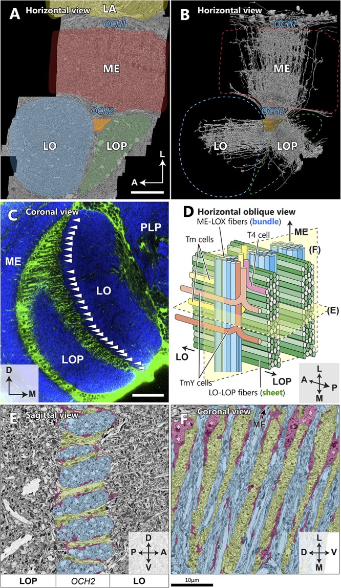
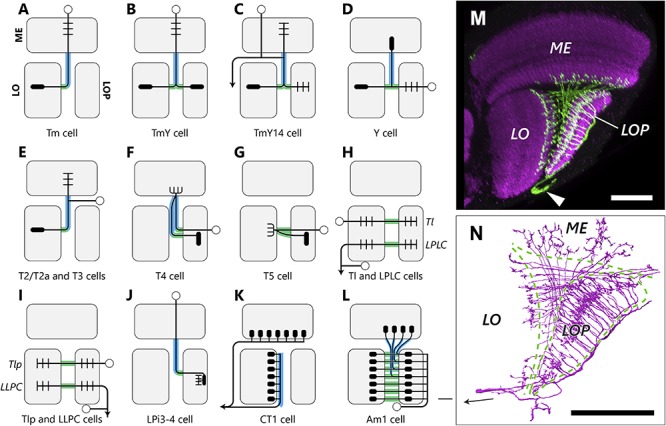
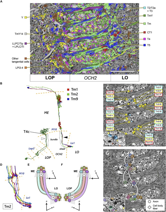
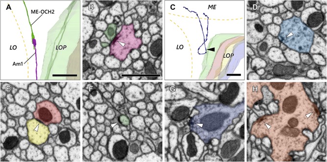
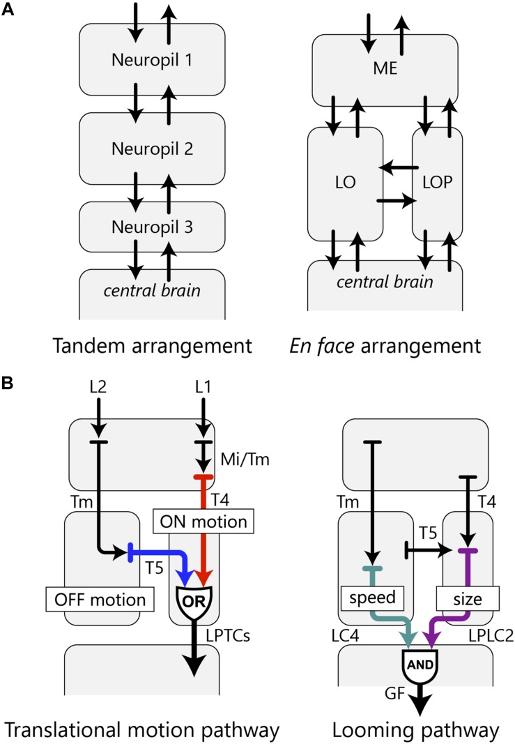
Visual pathways from the compound eye of an insect relay to four neuropils, successively the lamina, medulla, lobula, and lobula plate in the underlying optic lobe. Among these neuropils, the medulla, lobula, and lobula plate are interconnected by the complex second optic chiasm, through which the anteroposterior axis undergoes an inversion between the medulla and lobula. Given their complex structure, the projection patterns through the second optic chiasm have so far lacked critical analysis. By densely reconstructing axon trajectories using a volumetric scanning electron microscopy (SEM) technique, we reveal the three-dimensional structure of the second optic chiasm of Drosophila melanogaster, which comprises interleaving bundles and sheets of axons insulated from each other by glial sheaths. These axon bundles invert their horizontal sequence in passing between the medulla and lobula. Axons connecting the medulla and lobula plate are also bundled together with them but do not decussate the sequence of their horizontal positions. They interleave with sheets of projection neuron axons between the lobula and lobula plate, which also lack decussations. We estimate that approximately 19,500 cells per hemisphere, about two thirds of the optic lobe neurons, contribute to the second chiasm, most being Tm cells, with an estimated additional 2,780 T4 and T5 cells each. The chiasm mostly comprises axons and cell body fibers, but also a few synaptic elements. Based on our anatomical findings, we propose that a chiasmal structure between the neuropils is potentially advantageous for processing complex visual information in parallel. The EM reconstruction shows not only the structure of the chiasm in the adult brain, the previously unreported main topic of our study, but also suggest that the projection patterns of the neurons comprising the chiasm may be determined by the proliferation centers from which the neurons develop. Such a complex wiring pattern could, we suggest, only have arisen in several evolutionary steps.

Keywords: Drosophila melanogaster; lobula; lobula plate; medulla; optic chiasm; optic lobe; scanning electron microscopy; visual system.


Figures

FIGURE 1
FIGURE 1
Structure of the optic neuropils and their chiasmata. (A) Dorsal view of a fly’s head. The optic lobe forms a large part of the fly’s brain, and is situated lateral to the central brain in the head capsule, just beneath the retina of the compound eye. (B) A horizontal schematic view of the optic lobe and the topological projection patterns between the neuropils. The optic lobe comprises four stratified neuropils; the lamina (LA), medulla (ME), lobula (LO), and lobula plate (LOP). The outermost retina (RE) houses ommatidial clusters of photoreceptors that project directly to the lamina. Between the lamina and the medulla, the antero-posterior (A-P) retinotopic axis inverts via the first optic chiasm, OCH1, with the axons of lamina neurons decussating in the middle of the chiasm. Neurons corresponding to the posterior part of the visual field innervate the anterior part of the medulla. From there, neurons relaying the information project to the deepest (proximal) part of the lobula and lobula plate, traveling the longest path in the second optic chiasm, OCH2. Here, axons connecting the medulla and lobula decussate in the chiasm (red circle), while those between the medulla and lobula plate do not. Axons connecting the lobula and lobula plate run in parallel in the chiasm. A and P indicate anterior and posterior, respectively, both corresponding to the retinotopic orientation of the visual field. Names of representative layers according to Fischbach and Dittrich (1989) are shown. (C–F) Schematic views of chiasmal fiber projection patterns between the neuropils depicting the projection of OCH1 fibers (C), and corresponding to the three projection patterns of OCH2 neurons (D–F).
FIGURE 2
FIGURE 2
Spatial orientation of the second optic chiasm. (A) A re-sliced cross section of the optic lobe imaged with FIB-SEM. The OCH2 region of interest (ROI) used for dense neuron annotation in this study is highlighted in orange. The ROI was set so that it covered 4 bundles and part of 5 sheets, and had a thickness along the L-M axis of 10.5 μm, and a volume of ∼3,870 μm3. (B) Skeletonized image of 1,225 columnar neurons (∼50% of all those we traced) which intersect the OCH2 ROI. Neurons were randomly selected for visualization. (C) A coronal section depicting glial processes in OCH2. Glial tissues (green) are labeled with repo-GAL4>GFP, while synaptic neuropils (blue) are labeled with anti-Brp (nc82 antibody). Arrowheads indicate the bundles of fibers surrounded by glial processes. PLP: posterior lateral protocerebrum. (D) A model of the bundles and sheets of OCH2. Fibers in the bundles (shown in blue), running vertically in the panel, connect the medulla and lobula complex, and those in the sheets (shown in green) connect the lobula and lobula plate. Note that axons between the medulla and lobula complex defasciculate from their bundles and merge into the sheets, before projecting to the lobula complex neuropils. The bundle therefore becomes thinner as it runs deeper into the OCH2. Examples of T4 (pink), Tm (yellow), and TmY (orange, brown) cells are in different colors. (E) Cross section image of the bundles and sheets, cut perpendicular to the bundles and in parallel to the sheets. The bundles are labeled in blue, the sheets in green, and major glial processes surrounding the structures in red for (E) and (F). (F) Cross section of the chiasm, cut in parallel to the bundles and perpendicular to the sheets in a plane rotated 90° from that in (E). Cell bodies of glia are indicated by asterisks. Scale bar: (A,C) 30 μm, (E,F) 10 μm. Axes shown in (A), (C), (E), and (F) are based on the body axis.
FIGURE 3
FIGURE 3
Types and projection patterns of OCH2 neurons. (A–L) Neuron types that contribute to OCH2, and their projection patterns. Open circles indicate cell bodies, arrows indicate projections to/from the central brain in (H), (I) and (K). Short lines indicate typical input (dendritic/postsynaptic) neurites, and filled ellipses indicate typical output (axonal/presynaptic) neurites. Neuron arbors shown are considerably simplified, and pre- and postsynaptic motifs almost always coexist at single neurites in actual neurons, so that input and output sites are therefore not strictly segregated. Lines shown in green and blue, respectively, correspond to neuronal fibers in the bundles and sheets. (M) The Am1 cell visualized with FLP-out. Arrowhead indicates the cell body. (N) To compare with (M), Am1 reconstructed from the FIB-SEM dataset. The arrow in (N) indicates the cell body fiber. Scale bar: (M,N) 30 μm.
FIGURE 4
FIGURE 4
Topology of neurons in OCH2 pathways. (A) Distribution of different cell types in a cross-section of OCH2. “Am1 + tangential cells” include Am1 and other tangential elements including those not contributing to the chiasm. Neurons with dense core vesicles (DCV) are not color-coded. See also Table 1 and Figure 2E. White dashed line demarcates the area of OCH2 in which neurons are densely traced and annotated. (B) Reconstructed Tm cells and a T4c cell. Three Tm cells, Tm1, Tm2, and Tm9, innervate the same columns both in the medulla (ME) and lobula (LO), but are not tightly bundled in the chiasm (arrowhead), see text. The T4 cell has two fibers in the chiasm, a cell body fiber and an axon connecting the medulla and lobula plate (LOP; in this case the third layer, Lop3). Fibers contributing to the bundle or sheet in the chiasm are so indicated. These fibers do not necessarily run next to each other in the chiasm either (white arrowhead). (C) Axons of the Tm1, Tm2, and Tm9 cells from seven columns in OCH2. The neurons are color-coded by neuron types. The inset shows an enlarged view of part of the chiasm, indicated by a dotted box. (D) Six reconstructed Tm2 cells, a typical one-per-column neuron, in the chiasm from M10 to the lobula terminals. (E,F) Projection patterns of columnar neurons connecting the medulla and lobula (E) and the medulla and lobula plate (F). The spatial layout of the neurons as well as the color code of (E) are the same as for the panel (D). (G) Distribution of the axons and cell body fibers of T4 cells in OCH2. Circles indicate the axons, and squares indicate the cell body fibers. Each pair of profiles having the same color and connected with a white line belongs to a single T4 cell. The inset shows an enlarged view of part the chiasm, indicated by the dotted rectangle. Scale bar: (A,C,G) 10 μm. D, dorsal, P, posterior.
FIGURE 5
FIGURE 5
Axon and synaptic ultrastructure in the chiasm. (A) An Am1 branch and a ME-OCH2 cell terminal, both terminating in OCH2. (B) The two cells shown in (A) form a synaptic contact within OCH2. The two sites of synaptic contact are revealed where the Am1 terminal (red), forms a T-bar ribbon (arrowhead), which is presynaptic, opposite the ME-OCH2 terminal (green) where it is postsynaptic. (C) Part of a DCV cell making a reverse turn in OCH2. Its ME part is incompletely reconstructed. (D) A synaptic site of the DCV cell shown in (C). The synaptic site corresponds to the point indicated by the arrowhead in (C). The DCV cell has a presynaptic T-bar (arrowhead) and provides input to a dyad of postsynaptic Tm cell axons. (E,F) Putative septate or adherens junctions in OCH2. (E) is a case between Tm cell axons, and (F) is a case between a Tm cell axon and glia. (G,H) Typical presynaptic T-bars found outside OCH2. (G) is an example of a Tm1 cell, while (H) is a Tm9 cell terminal, both in the first stratum of the lobula. Scale bar: (A,C): 10 μm; (B), (D–H): 1 μm.
FIGURE 6
FIGURE 6
OCH2 and the arrangement of the optic lobe neuropils. (A) A hypothetical tandem arrangement (left) compared with the actual en face arrangement (right) of the optic lobe neuropils. The en face arrangement has more possible pathways between the neuropils as well as between the optic lobe and the central brain. (B) Two examples of information integration in the fly’s visual system accomplished by parallel information processing in multiple neuropils. The translational motion-processing pathway implements an OR operation, whereas the looming pathway implements an AND operation with the optic lobe neurons.

References

    1. Ache J. M., Polsky J., Alghailani S., Parekh R., Breads P., Peek M. Y., et al. (2019). Neural basis for looming size and velocity encoding in the Drosophila giant fiber escape pathway. Curr. Biol. 29 1073.e4–1081.e4. 10.1016/j.cub.2019.01.079 - DOI - PubMed
    1. Apitz H., Salecker I. (2014). A challenge of numbers and diversity: neurogenesis in the Drosophila optic lobe. J. Neurogenet. 28 233–249. 10.3109/01677063.2014.922558 - DOI - PubMed
    1. Apitz H., Salecker I. (2015). A region-specific neurogenesis mode requires migratory progenitors in the Drosophila visual system. Nat. Neurosci. 18 46–55. 10.1038/nn.3896 - DOI - PMC - PubMed
    1. Apitz H., Salecker I. (2016). Retinal determination genes coordinate neuroepithelial specification and neurogenesis modes in the Drosophila optic lobe. Development 143 2431–2442. 10.1242/dev.135004 - DOI - PMC - PubMed
    1. Apitz H., Salecker I. (2018). Spatio-temporal relays control layer identity of direction-selective neuron subtypes in Drosophila. Nat. Commun. 9:2295. 10.1038/s41467-018-04592-z - DOI - PMC - PubMed

Publication types

MeSH terms

LinkOut - more resources