Review
. 2019 Oct 17:10:2390.
doi: 10.3389/fimmu.2019.02390. eCollection 2019.

Bacterial Manipulation of Wnt Signaling: A Host-Pathogen Tug-of-Wnt

Affiliations
Review

Bacterial Manipulation of Wnt Signaling: A Host-Pathogen Tug-of-Wnt

Madison R Rogan et al. Front Immunol. .

Abstract

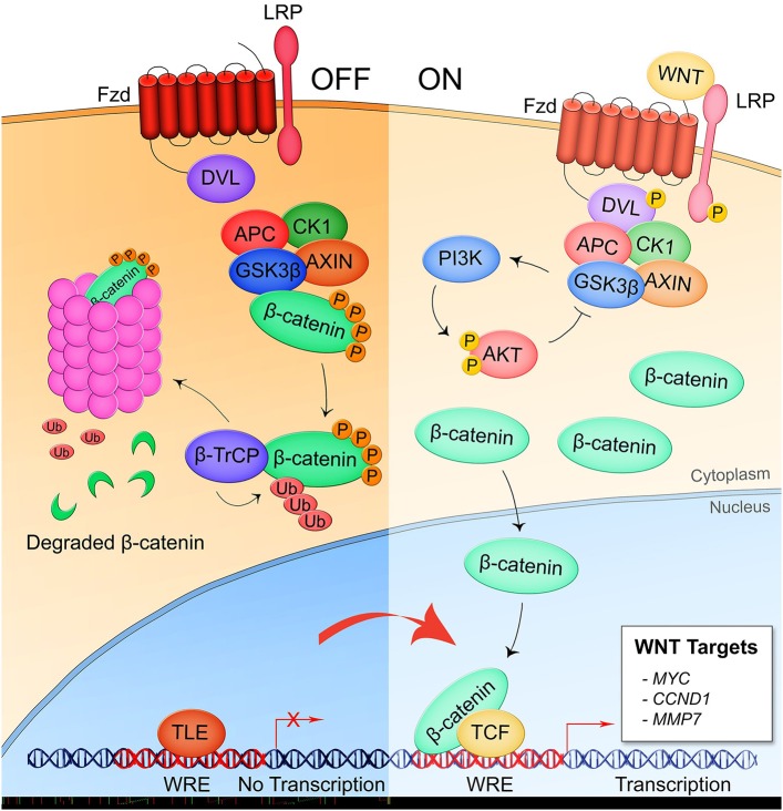
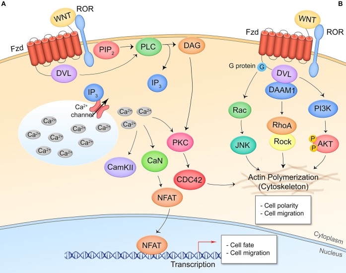
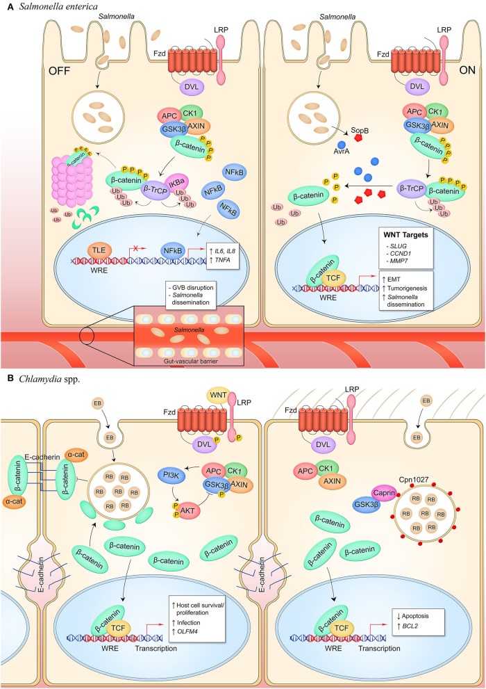
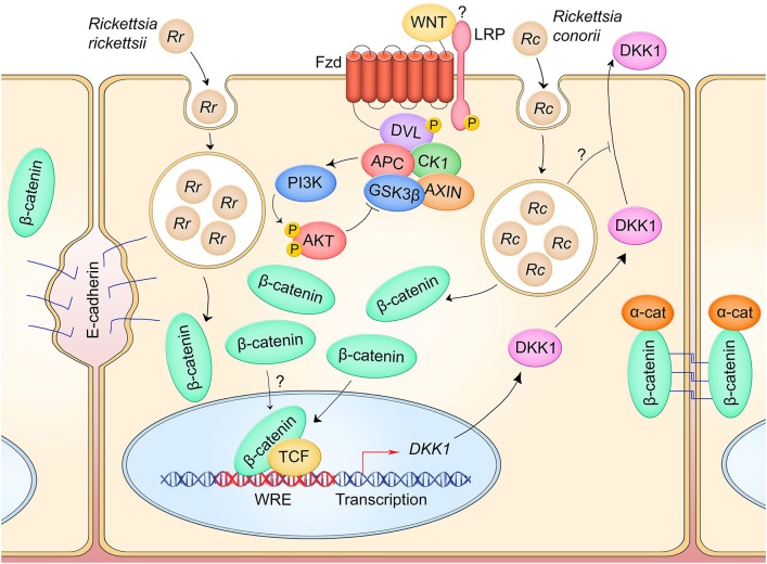
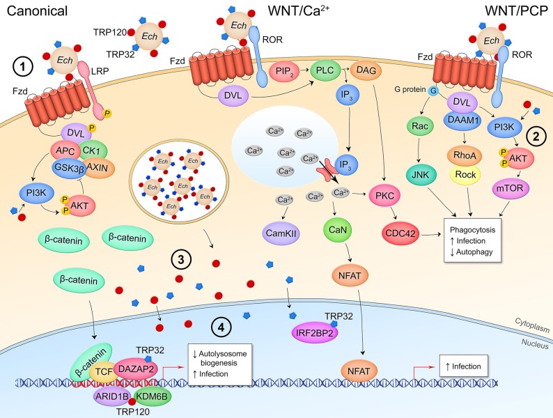
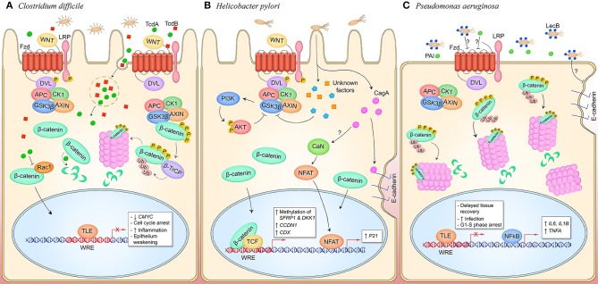
The host-pathogen interface is a crucial battleground during bacterial infection in which host defenses are met with an array of bacterial counter-mechanisms whereby the invader aims to make the host environment more favorable to survival and dissemination. Interestingly, the eukaryotic Wnt signaling pathway has emerged as a key player in the host and pathogen tug-of-war. Although studied for decades as a regulator of embryogenesis, stem cell maintenance, bone formation, and organogenesis, Wnt signaling has recently been shown to control processes related to bacterial infection in the human host. Wnt signaling pathways contribute to cell cycle control, cytoskeleton reorganization during phagocytosis and cell migration, autophagy, apoptosis, and a number of inflammation-related events. Unsurprisingly, bacterial pathogens have evolved strategies to manipulate these Wnt-associated processes in order to enhance infection and survival within the human host. In this review, we examine the different ways human bacterial pathogens with distinct host cell tropisms and lifestyles exploit Wnt signaling for infection and address the potential of harnessing Wnt-related mechanisms to combat infectious disease.

Keywords: Wnt; bacteria; immunoevasion; innate immunity; pathogen; β-catenin.


Figures

Figure 1
Figure 1
Canonical/β-catenin-dependent Wnt signaling. In the pathway off state, the β-catenin destruction complex consisting of APC, CK1, GSK3β, and AXIN binds β-catenin leading to its phosphorylation by GSK3β and subsequent ubiquitination by the E3 ubiquitin ligase complex β-TrCP which targets β-catenin for proteasomal degradation. Thus, the Wnt response element (WRE) located in the promoter region of Wnt pathway target genes remains bound by transcriptional repressor TLE. When a secreted Wnt ligand originating from the same or a nearby cell binds Fzd and the coreceptor LRP5/6, Dvl is phosphorylated and sequesters the destruction complex, preventing the phosphorylation of β-catenin. Accumulation of β-catenin is the cytoplasm leads to nuclear translocation of the protein where it binds with co-activator TCF at the WRE and drives expression of Wnt target genes.
Figure 2
Figure 2
Non-canonical/β-catenin-independent Wnt signaling pathways. (A) Wnt/Ca2+ signaling. A Wnt ligand interaction with Fzd and the non-canonical coreceptors ROR1/2 activates Dvl which activates PLC, leading to production of IP3 and opening of cytosolic Ca2+ stores. CAMKII, CaN, and PKC are activated by the increased cytosolic Ca2+ levels. CaN dephosphorylates NFAT leading to its nuclear translocation and expression of NFAT target genes. PKC crosstalks with Wnt/PCP signaling. (B) Wnt/planar cell polarity signaling. Interaction of Wnt and respective receptor/coreceptor leads to cytoskeletal reorganization through G-protein activation of Ras-JNK signaling, Dvl-DAAM1 association and Rho activation, and Dvl activation of PI3K and Akt. This pathway is transcription-independent.
Figure 3
Figure 3
Canonical Wnt signaling manipulation during bacterial infection of epithelial cells. (A) S. enterica infection of intestinal epithelial cells inhibits Wnt signaling through an unknown mechanism (left). Activity of the E3 ubiquitin ligase β-TrCP, which targets both β-catenin and IκBα for proteasomal degradation, stabilizes NF-κB levels which causes nuclear translocation of the protein and expression of pro-inflammatory target genes. Wnt signaling is also inhibited in the GVB during infection, promoting bacterial access to vasculature by increasing vascular permeability. S. enterica secretes the T3SS effectors AvrA and SopB into intestinal M cells and crypt-localized epithelial cells which causes activation of canonical Wnt signaling (right). Deubiquitinase AvrA and phosphatase SopB induce pathway activation through reversal of β-catenin posttranslational modifications, promoting expression of β-catenin-dependent genes that drive EMT in M cells, induce intestinal stem cell proliferation, and inhibit NF-κB activity in stem cells. (B) C. trachomatis infection of reproductive tract epithelium induces breakdown of adherens junctions and accumulation of β-catenin in the cytoplasm (left). β-catenin localizes to the chlamydial inclusion and translocates into the nucleus to activate transcription. Pathway activity promotes the bacterial developmental cycle through unknown mechanisms as well as drives expression of OLFM4 which is known to inhibit NF-κB signaling. C. pneumoniae expresses inclusion membrane protein Cpn1027 during infection of respiratory epithelium (right). Cpn1027 recruits Caprin2, a scaffold protein of the β-catenin destruction complex, as well as GSK3β, thereby reducing β-catenin turnover and allowing nuclear translocation for expression of target gene BCL2 to inhibit host cell apoptosis.
Figure 4
Figure 4
Canonical Wnt signaling manipulation during Rickettsia spp. infection of endothelium. R. rickettsii (Rr) infection of endothelial cells induces breakdown of adherens junctions and accumulation of cytoplasmic β-catenin which may drive pathway activity (left). R. conorii (Rc) infection leads to a suppression of secreted Wnt antagonist DKK1, which may facilitate activation of Wnt signaling to induce an anti-inflammatory environment during infection. IL-6 and IL-8 secretion are suppressed during infection in a DKK1-dependent manner.
Figure 5
Figure 5
Canonical and non-canonical Wnt signaling manipulation during E. chaffeensis infection of monocytes. (1) E. chaffeensis dense-cored cells express surface proteins TRP32 and TRP120 that stimulate phagocytosis in a Wnt signaling-dependent manner, potentially through interaction with the Wnt receptor complex and stimulation of non-canonical Wnt pathways to induce cytosolic Ca2+ flux and cytoskeletal reorganization. (2) Pathway activity during bacterial entry as well as T1SS secreted effectors TRP32 and TRP120 direct stimulation of PI3K-AKT signaling facilitates mTOR activation which prevents fusion of the bacterial replicative autophagosome with the lysosome. During infection, the secreted TRP effectors localize to the host cytoplasm where they interact with proteins involved in Wnt signaling (3) as well as translocate to the nucleus where they bind host DNA within the promoter regions of Wnt target genes or directly interact with transcriptional regulators of Wnt target genes (4).
Figure 6
Figure 6
Canonical and non-canonical Wnt signaling manipulation by pathogens occupying an extracellular niche. (A) C. difficile secretes toxins TcdA and TcdB which are phagocytosed by host cells in the intestinal and colonic epithelia. TcdA glucosyltransferase activity inhibits small GTPases like RacA which may inhibit pathway activity through inhibiting nuclear translocation of β-catenin. TcdB interacts with Wnt pathway components LRP6, Wnt5a, and GSK3β, and Fzd1, 2, and 7. Direct binding of Fzds prevents binding of endogenous Wnt ligands and silences pathway activity. This promotes epithelium permeability and a pro-inflammatory state. (B) During H. pylori infection of gastric mucosa, CagA activates canonical Wnt signaling through the breakdown of adherens junctions, and non-canonical Wnt signaling through stimulation of CaN and NFAT nuclear translocation by an unknown mechanism. Canonical pathway signaling is also activated through unknown T4SS effectors that induce phosphorylation of LRP and DVL and inhibit GSK3β, promoting cytosolic accumulation of β-catenin and subsequent nuclear translocation to activate genes that drive cell proliferation. Furthermore, unknown mechanisms induce methylation of Wnt pathway inhibitory genes SFRP1 and DKK1 to hypothetically facilitate Wnt signaling activity during infection. (C) P. aeruginosa is decorated with lectin LecB which induces β-catenin degradation through an unknown extracellular mechanism, resulting in decreased cell proliferation. Coincidentally, NF-κB signaling is activated and inflammatory target genes IL6, IL1B, and TNFA are expressed. LecB as well as the quorum-sensing molecule PAI both induce disruption of adherens junction in infected epithelia, contributing to prolonged tissue damage in the host.

References

    1. Christophides GK, Zdobnov E, Barillas-Mury C, Birney E, Blandin S, Blass C, et al. . Immunity-related genes and gene families in Anopheles gambiae. Science. (2002) 298:159–65. 10.1126/science.1077136 - DOI - PubMed
    1. Mogensen TH. Pathogen recognition and inflammatory signaling in innate immune defenses. Clin Microbiol Rev. (2009) 22:240–73. 10.1128/CMR.00046-08 - DOI - PMC - PubMed
    1. Takeuchi O, Akira S. Pattern recognition receptors and inflammation. Cell. (2010) 140:805–20. 10.1016/j.cell.2010.01.022 - DOI - PubMed
    1. Riera Romo M, Pérez-Martínez D, Castillo Ferrer C. Innate immunity in vertebrates: an overview. Immunology. (2016) 148:125–39. 10.1111/imm.12597 - DOI - PMC - PubMed
    1. Siebel C, Lendahl U. Notch signaling in development, tissue homeostasis, and disease. Physiol Rev. (2017) 97:1235–94. 10.1152/physrev.00005.2017 - DOI - PubMed

Publication types

MeSH terms