

Excitation, but not inhibition, of the fastigial nucleus provides powerful control over temporal lobe seizures
- PMID: 31682010
- PMCID: PMC6938547
- DOI: 10.1113/JP278747
Excitation, but not inhibition, of the fastigial nucleus provides powerful control over temporal lobe seizures
Abstract
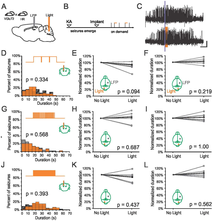
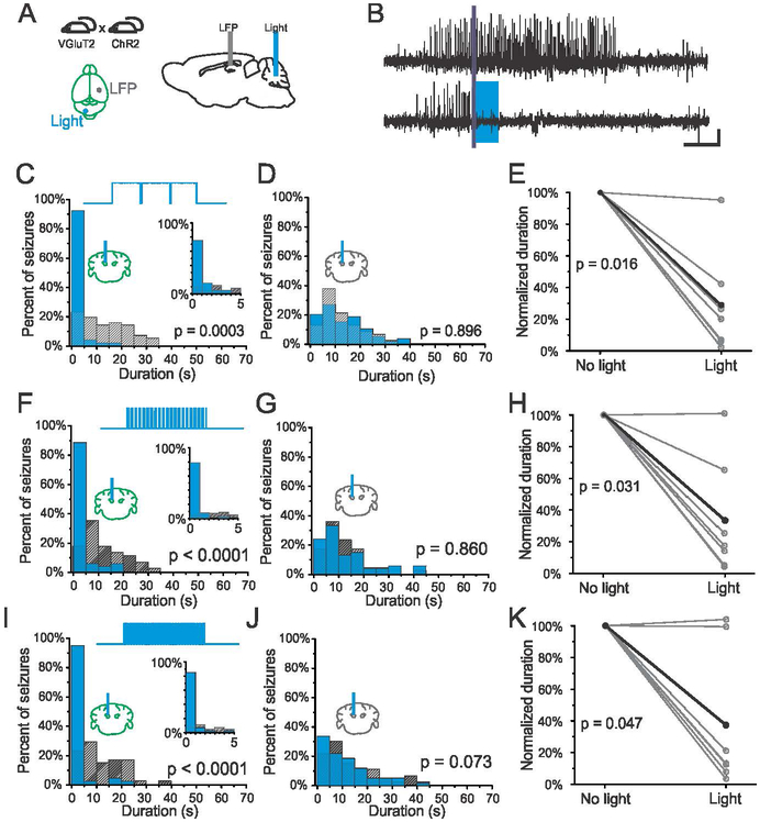
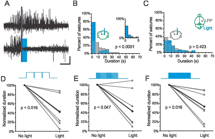
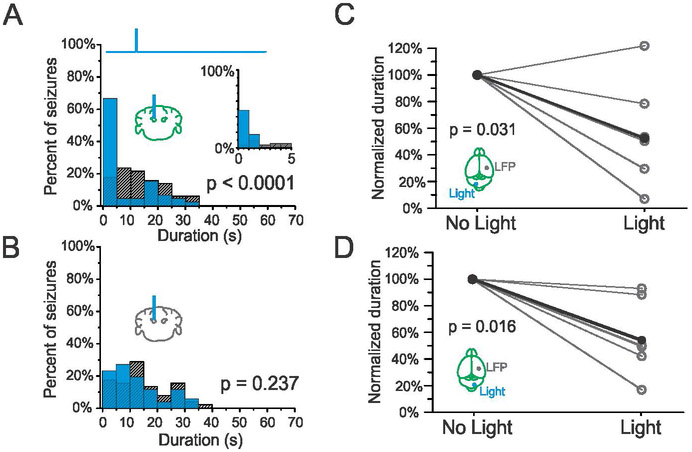
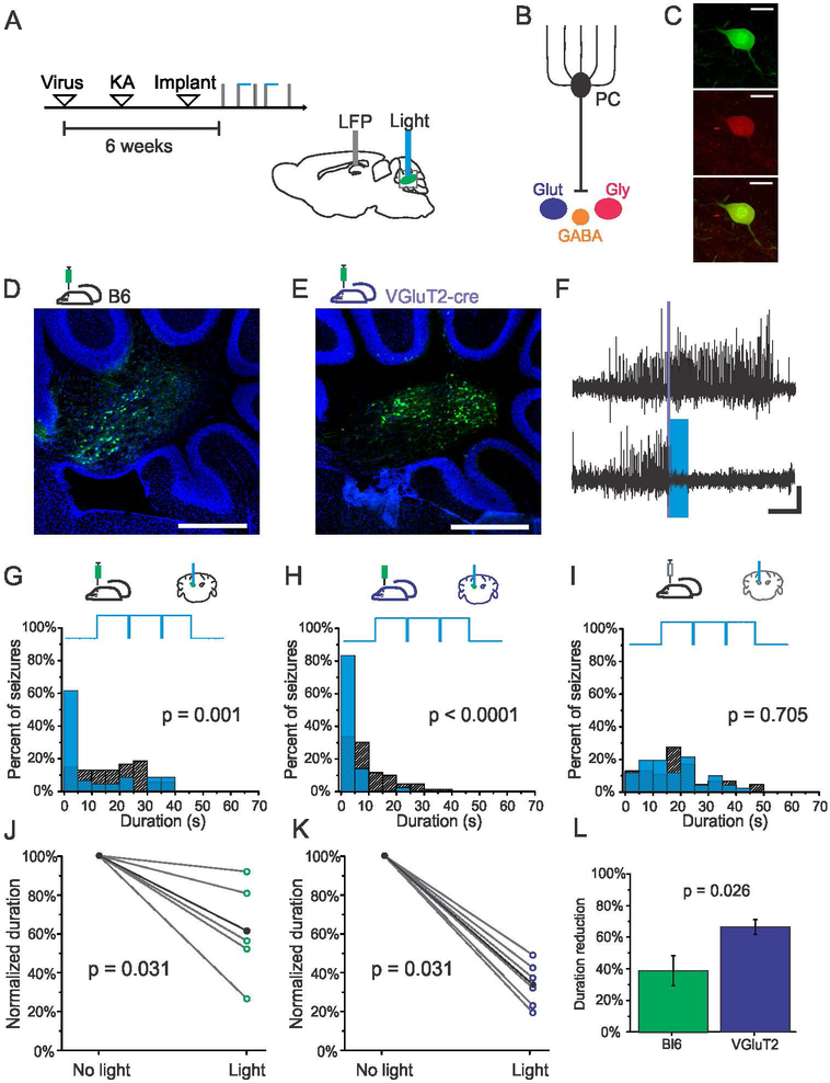
Key points: On-demand optogenetic inhibition of glutamatergic neurons in the fastigial nucleus of the cerebellum does not alter hippocampal seizures in a mouse model of temporal lobe epilepsy. In contrast, on-demand optogenetic excitation of glutamatergic neurons in the fastigial nucleus successfully inhibits hippocampal seizures. With this approach, even a single 50 ms pulse of light is able to significantly inhibit seizures. On-demand optogenetic excitation of glutamatergic fastigial neurons either ipsilateral or contralateral to the seizure focus is able to inhibit seizures. Selective excitation of glutamatergic nuclear neurons provides greater seizure inhibition than broadly exciting nuclear neurons without cell-type specificity.
Abstract: Temporal lobe epilepsy is the most common form of epilepsy in adults, but current treatment options provide limited efficacy, leaving as many as one-third of patients with uncontrolled seizures. Recently, attention has shifted towards more closed-loop therapies for seizure control, and on-demand optogenetic modulation of the cerebellar cortex was shown to be highly effective at attenuating hippocampal seizures. Intriguingly, both optogenetic excitation and inhibition of cerebellar cortical output neurons, Purkinje cells, attenuated seizures. The mechanisms by which the cerebellum impacts seizures, however, are unknown. In the present study, we targeted the immediate downstream projection of vermal Purkinje cells - the fastigial nucleus - in order to determine whether increases and/or decreases in fastigial output can underlie seizure cessation. Though Purkinje cell input to fastigial neurons is inhibitory, direct optogenetic inhibition of the fastigial nucleus had no effect on seizure duration. Conversely, however, fastigial excitation robustly attenuated hippocampal seizures. Seizure cessation was achieved at multiple stimulation frequencies, regardless of laterality relative to seizure focus, and even with single light pulses. Seizure inhibition was greater when selectively targeting glutamatergic fastigial neurons than when an approach that lacked cell-type specificity was used. Together, these results suggest that stimulating excitatory neurons in the fastigial nucleus may be a promising approach for therapeutic intervention in temporal lobe epilepsy.
Keywords: cerebellum; fastigial nucleus; optogenetics; temporal lobe epilepsy.
© 2019 The Authors. The Journal of Physiology © 2019 The Physiological Society.
Figures
Similar articles
-
Distinct Fastigial Output Channels and Their Impact on Temporal Lobe Seizures.J Neurosci. 2021 Dec 8;41(49):10091-10107. doi: 10.1523/JNEUROSCI.0683-21.2021. Epub 2021 Oct 29. J Neurosci. 2021. PMID: 34716233 Free PMC article.
-
Cerebellar Directed Optogenetic Intervention Inhibits Spontaneous Hippocampal Seizures in a Mouse Model of Temporal Lobe Epilepsy.eNeuro. 2014 Dec;1(1):ENEURO.0005-14.2014. doi: 10.1523/ENEURO.0005-14.2014. eNeuro. 2014. PMID: 25599088 Free PMC article.
-
Medial septal GABAergic neurons reduce seizure duration upon optogenetic closed-loop stimulation.Brain. 2021 Jun 22;144(5):1576-1589. doi: 10.1093/brain/awab042. Brain. 2021. PMID: 33769452 Free PMC article.
-
The cerebellum and epilepsy.Epilepsy Behav. 2021 Aug;121(Pt B):106909. doi: 10.1016/j.yebeh.2020.106909. Epub 2020 Feb 5. Epilepsy Behav. 2021. PMID: 32035793 Free PMC article. Review.
-
Optogenetics for controlling seizure circuits for translational approaches.Neurobiol Dis. 2023 Aug;184:106234. doi: 10.1016/j.nbd.2023.106234. Epub 2023 Jul 20. Neurobiol Dis. 2023. PMID: 37479090 Review.
Cited by
-
Pilot Safety and Feasibility Study of Non-invasive Limb Proprioceptive Cerebellar Stimulation for Epilepsy.Front Neurol. 2021 Aug 17;12:675947. doi: 10.3389/fneur.2021.675947. eCollection 2021. Front Neurol. 2021. PMID: 34484096 Free PMC article.
-
Highlights From AES2020, a Virtual American Epilepsy Society Experience.Epilepsy Curr. 2021 May 16;21(4):15357597211018219. doi: 10.1177/15357597211018219. Online ahead of print. Epilepsy Curr. 2021. PMID: 33998298 Free PMC article.
-
Individual cerebellar metabolic connectome in patients with MTLE and NTLE associated with surgical prognosis.Eur J Nucl Med Mol Imaging. 2024 Oct;51(12):3600-3616. doi: 10.1007/s00259-024-06762-2. Epub 2024 May 28. Eur J Nucl Med Mol Imaging. 2024. PMID: 38805089
-
Mapping Lesion-Related Epilepsy to a Human Brain Network.JAMA Neurol. 2023 Sep 1;80(9):891-902. doi: 10.1001/jamaneurol.2023.1988. JAMA Neurol. 2023. PMID: 37399040 Free PMC article.
-
LINCs Are Vulnerable to Epileptic Insult and Fail to Provide Seizure Control via On-Demand Activation.eNeuro. 2023 Feb 15;10(2):ENEURO.0195-22.2022. doi: 10.1523/ENEURO.0195-22.2022. Print 2023 Feb. eNeuro. 2023. PMID: 36725340 Free PMC article.
References
-
- Angaut P & Bowsher D. (1970). Ascending projections of the medial cerebellar (fastigial) nucleus: an experimental study in the cat. Brain Res 24, 49–68. - PubMed
-
- Babb TL, Mitchell AG Jr. & LCrandall PH. (1974). Fastigiobulbar and dentatothalamic influences on hippocampal cobalt epilepsy in the cat. Electroencephalogr Clin Neurophysiol 36, 141–154. - PubMed
Publication types
MeSH terms
Grants and funding
LinkOut - more resources
Full Text Sources
Medical
Molecular Biology Databases