. 2019 Nov 4;14(11):e0223800.
doi: 10.1371/journal.pone.0223800. eCollection 2019.

Protective effect of Platymiscium floribundum Vog. in tree extract on periodontitis inflammation in rats

Affiliations

Protective effect of Platymiscium floribundum Vog. in tree extract on periodontitis inflammation in rats

Jordânia M O Freire et al. PLoS One. .

Abstract

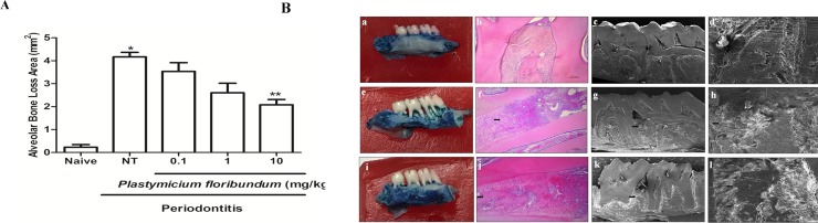
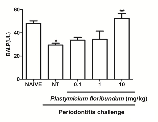
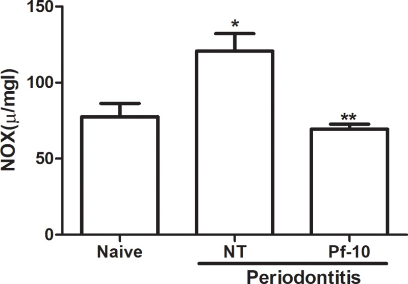
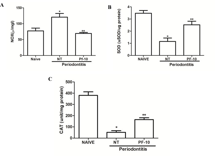
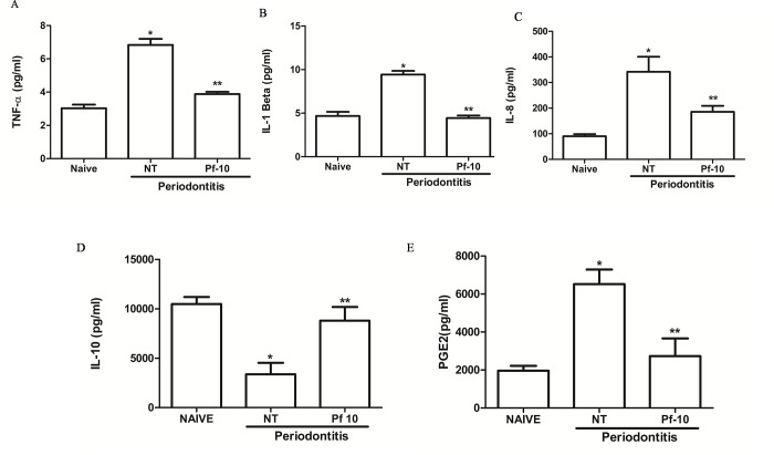
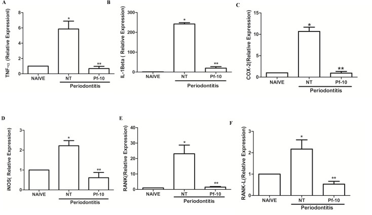
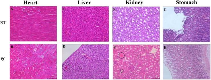
Periodontitis is an immuno-inflammatory disease, which can lead to tooth loss. This study aimed to investigate the efficacy of Platymiscium floribundum Vog., a Brazilian tree which has been used in folk medicine as an anti-inflammatory agent, in a pre-clinical trial of periodontitis in rats. Periodontitis was induced by placing a sterilized nylon (3.0) thread ligature around the cervix of the second left upper molar of the rats, which received (per os) P. floribundum extract (0.1, 1 or 10 mg/kg) or vehicle 1h before periodontitis-challenge and once daily during 11 days. Treatment with P. floribundum (10mg/kg) decreased alveolar bone loss, MPO activity nitrite/nitrate levels, oxidative stress, TNF-α, IL1-β, IL-8/CINC-1, and PGE2 gingival levels, and transcription of TNF-α, IL1-β, COX-2, iNOS, RANK, and RANKL genes, while elevated both BALP serum levels and IL-10 gingival levels. The animals did not show signs of toxicity throughout the experimental course. These findings show that P. floribundum has anti-inflammatory and anti-resorptive properties in a pre-clinical trial of periodontitis, representing an interesting biotechnological tool.


Conflict of interest statement

The authors have declared that no competing interests exist.

Figures

Fig 1
Fig 1
(A) Effect of P. floribundum (0.1; 1 or 10 mg/kg) on alveolar bone loss in experimental periodontitis. Naive: animals without periodontitis; NT: animals subjected to periodontitis and treated with vehicle (0.9% saline + 0.1% ethanol). P. floribundum: rats submitted to periodontitis and treated with P. floribundum (0.1; 1 or 10 mg/kg), respectively. Data are shown as mean ± SEM (n = 6 for each treatment). *P < 0.000039 versus naive; **P < 0.001 versus NT, (ANOVA; Games-Howell). (B) Effect of P. floribundum (0.1; 1 or 10 mg/kg) on the macroscopic view (first column), histological aspects (second column), and Scanning Electron Microscopy (SEM) (third columm) of periodontium. (a–d) normal maxilla (naive), showing the integrity of its components (C—Cementum; D–Dentin, and AB—Alveolar Bone). (e–h) maxilla from rats subjected to periodontitis and receiving only vehicle (0.9% saline + 0.1% ethanol) presenting severe bone resorption, inflammatory infiltrate in the gingiva and periodontal ligament, extensive destruction of cementum, total resorption of the alveolar process (f), and irregularity in the bone tissue (g—h). (i–l) maxilla from rats subjected to periodontitis and treated with P. floribundum (10 mg/kg) showing discrete cellular influx and preservation of cementum and alveolar process (j) and regular tissue topography (k–l). Black arrows indicate alveolar bone resorption. HE magnification (100x); MEV magnification 65x: c, g, and k; MEV magnification 350x: d, h, and i.
Fig 2
Fig 2. Effect of P. floribundum on the serum alkaline phosphatase levels.
Data represent the mean ± SEM of six animals for each group. *p<0.001 versus naive; **p<0.012 versus non-tretaed (NT) (ANOVA; post hoc Games-Howell).
Fig 3
Fig 3. Effect of P. floribundum (10mg/kg) on gingival myeloperoxidase (MPO) activity.
Data are shown as mean ± SEM (n = 6 for each treatment). *P < 0.000023 versus naive; **P < 0.01046 versus non-treated NT, (ANOVA; Tukey).
Fig 4
Fig 4
(A) Effect of P. floribundum (10mg/kg) on gingival nitrite/nitrate levels (NOx). Data are shown as mean ± SEM (n = 6 for each treatment). *P < 0. 0015 versus naive; **P < 0. 0317 versus non-treated (NT) (ANOVA; Tukey). (B) Effect of P. floribundum (10mg/kg) on gingival SOD (B) and CAT (C) levels. Data are shown as mean ± SEM (n = 6 for each treatment). * P < 0.0003 versus naive; ** p< 0.013 versus NT (ANOVA; Tukey). *P < 0.007 versus naive; **P < 0.002 versus non-treated (NT) (ANOVA; Games-Howell).
Fig 5
Fig 5
Effects of P. floribundum on TNF-α (A), IL-1β (B), IL-8/CINC-1 (C), IL-10 (D), and PGE2 levels in gingival tissues. Data are shown as mean ± SEM (n = 6 for each treatment). *P < 0.00001 versus Naive and **P < 0.001 versus NT for TNF-α; *P < 0.017 versus Naive and **P < 0.05 versus NT for IL-8; *P < 0.000001 versus Naive and **P < 0.0000001 versus NT for IL-1β; *P < 0.000 versus Naive and **P < 0.004 versus NT for PGE2; *P < 0,01 versus Naive and **P < 0.02 versus NT for IL-10 (ANOVA; Games-Howell; post hoc Tukey).
Fig 6
Fig 6. Effect of P. floribundum (10mg/kg) on the mRNA levels of TNF-α, IL-1β, COX-2, iNOS, RANK, and RANK-L in gingival tissues from rats subjected to periodontitis.
Data are shown as mean ± SEM (n = 6 for each treatment). A. * P < 0.001 versus Naive; **P < 0.001 versus NT. B. *P < 0.0000001 versus Naive; **P < 0.0000001 versus NT. C. * P < 0.000004 versus Naive; **P < 0.000004 versus NT. D. * P < 0.007 versus Naive; ** P < 0.001 versus NT. E. *P < 0.036 versus Naive; ** P < 0.039 versus NT. F. * P < 0.05 versus Naive; ** P < 0.036 versus NT (ANOVA; Tukey; Games-Howell, respectively).
Fig 7
Fig 7. Photomicrographs of organs from rats subjected to periodontitis and treated with P. floribundum (10mg/kg).
(A) Heart, (C) Liver, (E) Kidney, and (G) Stomach from non-treated (NT) group (rats receiving only vehicle). (B) Heart, (D) Liver, (F) Kidney, and (H) Stomach from rats treated P. floribundum (10mg/kg). Black circles indicate cellular swelling, few hemorrhagic areas (D), areas of discrete congestion (F). Magnification 100x.

References

    1. Hajishengallis G. The inflammophilic character of the periodontitis‐associated microbiota. Mol Oral Microbiol. 2014; 29 (6): 248–257. 10.1111/omi.12065 - DOI - PMC - PubMed
    1. Trindade F, Oppenheim FG, Helmerhorst EJ, Amado F, Gomes PS, Vitorino R. Uncovering the molecular networks in periodontitis. Proteomics Clin Appl. 2014; 8 (9–10): 748–761. 10.1002/prca.201400028 - DOI - PMC - PubMed
    1. Sukhtankar L, Kulloli A, Kathariya R, Shetty S. Effect of non-surgical periodontal therapy on superoxide dismutase levels in gingival tissues of chronic periodontitis patients: A clinical and spectophotometric analysis. Dis Markers. 2013; 34 (5): 305–311. 10.3233/DMA-130978 - DOI - PMC - PubMed
    1. Uslu MÖ, Eltas A, Marakoğlu İ, Dündar S, Şahin K., Özercan İ H. Effects of diode laser application on inflammation and mpo in periodontal tissues in a rat model. J Appl Oral Sci. 2018; 26 (0): e20170266 10.1590/1678-7757-2017-0266 - DOI - PMC - PubMed
    1. Buommino E, Scognamiglio M, Donnarumma G, Fiorent A. Recent advances in natural product-based anti-biofilm approaches to control infections. Mini Rev Med Chem. 2014; 14 (14): 1169–1182. 10.2174/1389557515666150101095853 - DOI - PubMed

Publication types