Review
. 2020 Feb:60:7-16.
doi: 10.1016/j.sbi.2019.09.001. Epub 2019 Nov 1.

Amyloid structures: much more than just a cross-β fold

Affiliations
Review

Amyloid structures: much more than just a cross-β fold

Rodrigo Gallardo et al. Curr Opin Struct Biol. 2020 Feb.

Abstract

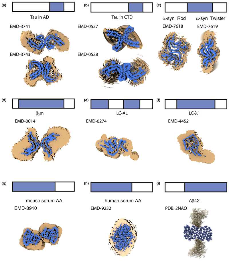
In recent years our understanding of amyloid structure has been revolutionised by innovations in cryo-electron microscopy, electron diffraction and solid-state NMR. These techniques have yielded high-resolution structures of fibrils isolated from patients with neurodegenerative disease, as well as those formed from amyloidogenic proteins in vitro. The results not only show the expected cross-β amyloid structure, but also reveal that the amyloid fold is unexpectedly diverse and complex. Here, we discuss this diversity, highlighting dynamic regions, ligand binding motifs, cavities, non-protein components, and structural polymorphism. Collectively, these variations combine to allow the generic amyloid fold to be realised in three dimensions in different ways, and this diversity may be related to the roles of fibrils in disease.


Conflict of interest statement

Conflict of interest statement

Nothing declared.

Figures

Figure 1
Figure 1. Updating the amyloid fold.
(a) Schematic of the cross-β fold viewed from (1) side and (2) cross-section, representing the stacking of molecular layers perpendicular to the long axes of the amyloid fibrils. (b) Detail of the structural elements observed in amyloid cores highlighting (1) in-register parallel β-sheets [•], (2) ‘dry’ steric zippers [29], (3) sidechain-mainchain loop hydrogen bonding [37], (4) polar zippers [26•], (5) buried salt-bridges [36••], (6) structured solvent molecules [••], (7) polar and apolar channels (green and purple arrows, respectively) [••], (8) cofactors (blue density) [••] and (9) multi-molecular layer interactions between central layer ‘L’ (blue) and the adjacent layers above and below (gold) [35].
Figure 2
Figure 2. The many faces of an amyloid.
Fibril polymorphs observed for (a) peptide fragments of LC λ [46] and for the full-length proteins (b) β2m [•], (c) Tau in AD [•], (d)40 [19,22], (e) Tau in Pick’s Disease [•], (f) α-synuclein [32], (g) mouse and human serum AA [35], (h)42 [24,29], (i) Tau in presence of heparin [38] and (j) segments of TDP43 [48]. These fibrils were isolated ex vivo (c,e,g) or formed in vitro (a,b,d,f,h,i,j). Illustrations in (a) and (b) are reproduced with permission [•,46].
Figure 3
Figure 3. Amyloid is more than a rigid core.
LDRs are shown on the structures of amyloid fibrils of full-length proteins formed in vitro or isolated ex vivo. The top of each panel shows a schematic of the full-length sequence (bar) with the sequence involved in forming the ordered amyloid core in blue and LDRs in white. The image on panels (a−h) corresponds to an orthogonal view down the fibril axis of the reported density maps contoured at two different levels. The regions of localised disorder are shown as broad/noisy density (orange) surrounding the amyloid core density (blue) for (a) Tau in Alzheimer’s disease [•], (b) Tau in CTE [••], (c) two polymorphs of α-synuclein [32], (d) β2m [•], (e) IGLV6-57 derived LC amyloid [37], (f) GLV1-44 derived LC amyloid [36••], (g) mouse AA and (h) human AA [35]. Panel (i) shows the structure of Aβ42 fibrils determined by ssNMR [24], where regions of disorder are modeled as loops that point away from the core. The EMD code of each map used is indicated on each panel. The blue maps are countered to the recommended level indicated for the deposited maps. The orange maps are 5 Å low-pass filtered of the deposited map countered to 1.75 RMS using ChimeraX [72].
Figure 4
Figure 4
A combination of techniques is required to understand the amyloid fibril structure and its cellular consequences. However, the picture is still incomplete. The missing aspects will be achieved through biochemical, biophysical and cellular investigation. Only by an integrative approach in which in vitro, ex vivo, in situ and in vivo approaches are combined can we hope to achieve the structural, cellular and mechanistic understanding required to fully understand the amyloid structure and to inspire biomedical progress.

Similar articles

Cited by

References

    1. Iadanza MG, Jackson MP, Hewitt EW, Ranson NA, Radford SE. A new era for understanding amyloid structures and disease. Nat Rev Mol Cell Biol. 2018;19:755–773. [Comprehensive review on the history of discovery, fibrillation and the relationship of fibril structure and disease] - PMC - PubMed
    1. Bowman GR, Voelz VA, Pande VS. Taming the complexity of protein folding. Curr Opin Struct Biol. 2011;21:4–11. doi: 10.1016/j.sbi.2010.10.006. - DOI - PMC - PubMed
    1. Babu MM, van der Lee R, de Groot NS, Gsponer J. Intrinsically disordered proteins: regulation and disease. Curr Opin Struct Biol. 2011;21:432–440. - PubMed
    1. Fernandez-Escamilla AM, Rousseau F, Schymkowitz J, Serrano L. Prediction of sequence-dependent and mutational effects on the aggregation of peptides and proteins. Nat Biotechnol. 2004;22:1302–1306. - PubMed
    1. Knowles TP, Waudby CA, Devlin GL, Cohen SI, Aguzzi A, Vendruscolo M, Terentjev EM, Welland ME, Dobson CM. An analytical solution to the kinetics of breakable filament assembly. Science. 2009;326:1533–1537. - PubMed

Publication types