Review
. 2020 Feb;40(1):84-90.
doi: 10.1055/s-0039-3399502. Epub 2019 Nov 4.

Mechanotransduction in Liver Diseases

Affiliations
Review

Mechanotransduction in Liver Diseases

Ningling Kang. Semin Liver Dis. 2020 Feb.

Abstract

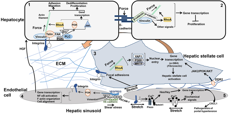
Chronic liver diseases, such as fibrosis and cancer, lead to a rigid or stiff liver because of perpetual activation of hepatic stellate cells or portal fibroblasts into matrix-producing myofibroblasts. Mechanical forces, as determined by the mechanical properties of extracellular matrix or pressure of circulating blood flow/shear stress, are sensed by mechanoreceptors at the plasma membrane and transmitted into a cell to impact cell function. This process is termed as mechanotransduction. This review includes basic knowledge regarding how external forces are sensed, amplified, and transmitted into the interior of a cell as far as the nucleus to regulate gene transcription and generate biological responses. It also reviews literatures to highlight the mechanisms by which mechanical forces in a normal or diseased liver influence the phenotype of hepatocytes, hepatic stellate cells, portal fibroblasts, and sinusoidal endothelial cells, and these cells in turn participate in the initiation and progression of liver diseases.


Conflict of interest statement

No conflict of interest exists.

Figures

Figure 1.
Figure 1.
Mechanotransduction in liver resident cells. Cell 1. ECM-mediated mechanical forces induce activation of integrin and the formation of FAs comprised of actin-binding proteins, such as talin and vinculin, and signaling molecules, such as FAK, Src, PI3K and so on. Through a direct force transfer to the actin filaments and RhoA-mediated biochemical signaling, external forces are translated into actomyosin contractility, cytoskeleton remodeling, and gene transcription, culminating in a new phenotype of the cell. Cell 2. Forces on E-cadherin induce the formation of cadherin complexes, which transmit forces into the interior of the cell by the actin filaments and signaling molecule such as RhoA. E-cadherin-mediated signaling is essential for the development of contact inhibition of cell proliferation of epithelial cells. Cell 3. In HSCs, ECM-mediated forces or stretch of the plasma membrane activate integrin and its downstream signals leading to nuclear translocation of YAP1, p300 or MRTF, which subsequently turns on gene transcription for HSC activation. In addition, LSEC-dependent angiogenesis at the early-stage of fibrosis leads to ECM remodeling and mechanical forces that promote HSC activation by a collagen-DDR2/JAK2/PI3K/AKT mechanosignaling. Cell 4. Shear stress induces the formation of a mechanosensory protein complex comprised of PECAM-1, VE-cadherin, and VEGFR2. In this complex, VE-cadherin functions as an adaptor and PECAM-1 activates Src and binds to the intermediate vimentin filaments for force transmission. VEGFR2 activates PI3K, which leads to subsequent integrin activation and the biological responses, such as cell alignment in laminar shear and activation of NF-κB. In addition, shear stress-induced mechanosignaling leads to the release of hepatic growth factor (HGF) that triggers the proliferation and survival of adjacent hepatocytes. Cell 5. Mechanical stretch of the cell membrane of LSECs activates cation channels. In addition, pulsatile forces on LSECs, as a result of hepatic congestion, activate integrin/Piezo/Notch to induce the release of CXCL1 that participates in the pathogenesis of portal hypertension.

References

    1. Wells RG. The role of matrix stiffness in regulating cell behavior. Hepatology. 2008;47(4):1394–1400. - PubMed
    1. Venkatesh SK, Ehman RL. Magnetic resonance elastography of liver. Magn Reson Imaging Clin N Am. 2014;22(3):433–446. - PMC - PubMed
    1. Ferraioli G, Tinelli C, Dal Bello B, et al. Accuracy of real-time shear wave elastography for assessing liver fibrosis in chronic hepatitis C: a pilot study. Hepatology. 2012;56(6):2125–2133. - PubMed
    1. Friedman SL. Mechanisms of disease: Mechanisms of hepatic fibrosis and therapeutic implications. Nat Clin Pract Gastroenterol Hepatol. 2004;1(2):98–105. - PubMed
    1. Olsen AL, Bloomer SA, Chan EP, et al. Hepatic stellate cells require a stiff environment for myofibroblastic differentiation. Am J Physiol Gastrointest Liver Physiol. 2011;301(1):G110–118. - PMC - PubMed

Publication types