. 2019 Oct 31;9(11):302.
doi: 10.3390/brainsci9110302.

D-Stellate Neurons of the Ventral Cochlear Nucleus Decrease in Auditory Nerve-Evoked Activity during Age-Related Hearing Loss

Affiliations

D-Stellate Neurons of the Ventral Cochlear Nucleus Decrease in Auditory Nerve-Evoked Activity during Age-Related Hearing Loss

Yong Wang et al. Brain Sci. .

Abstract

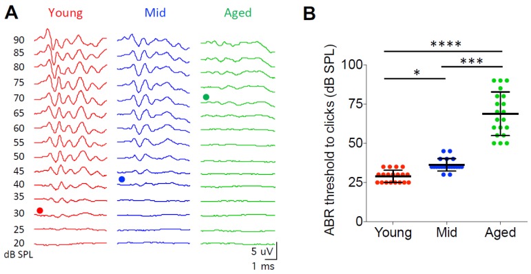
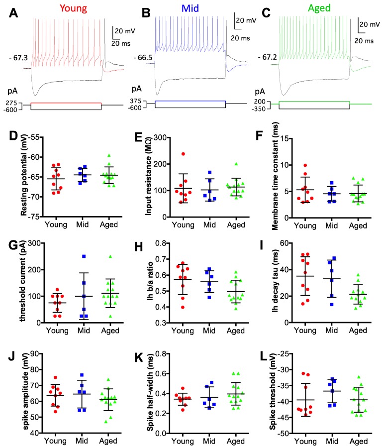
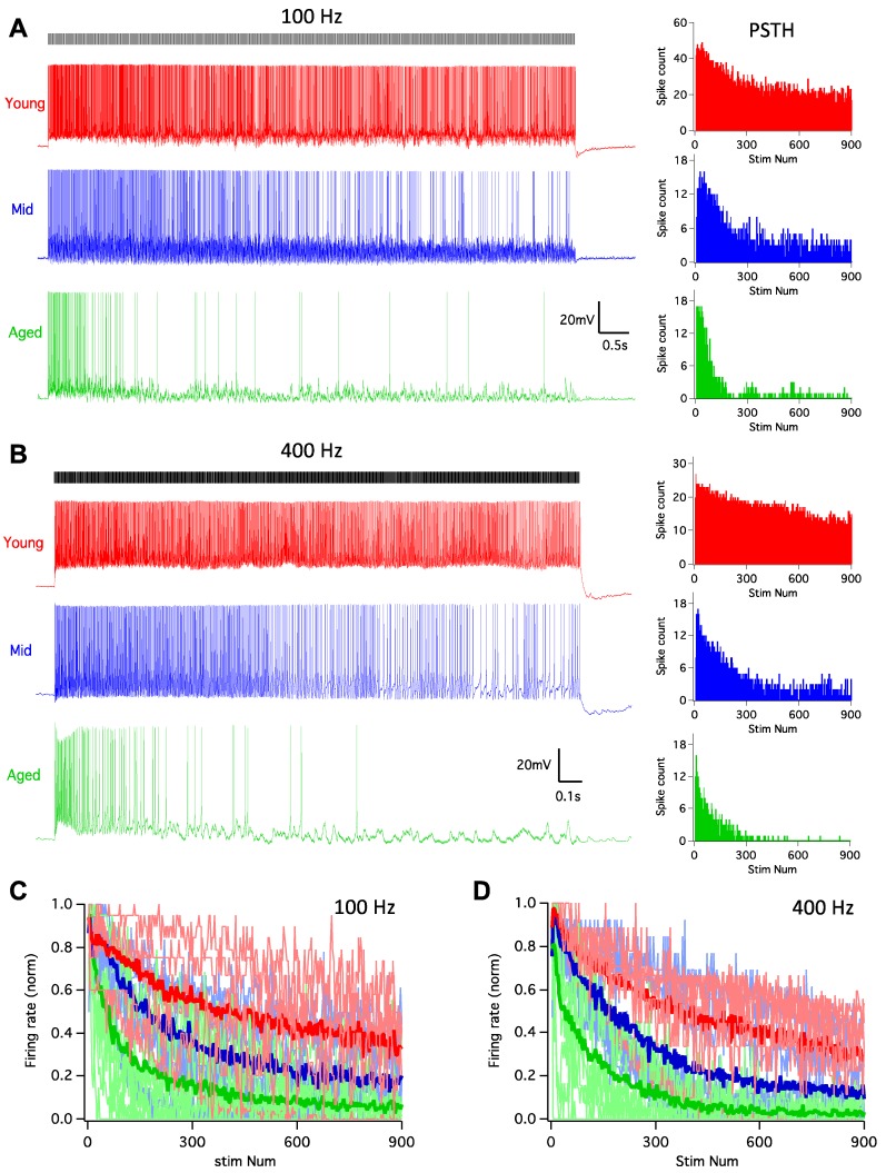
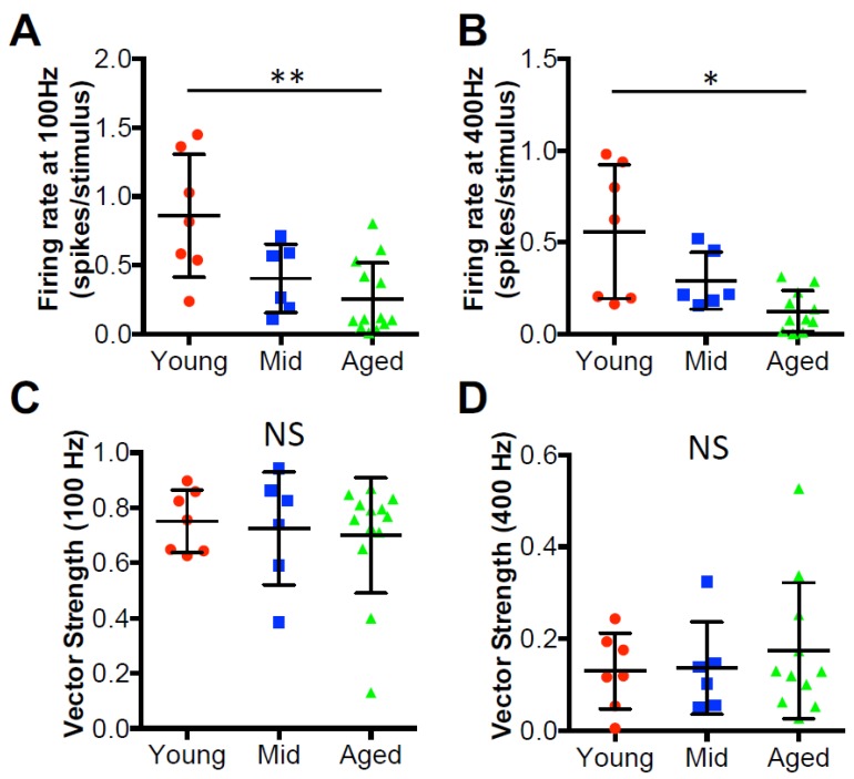
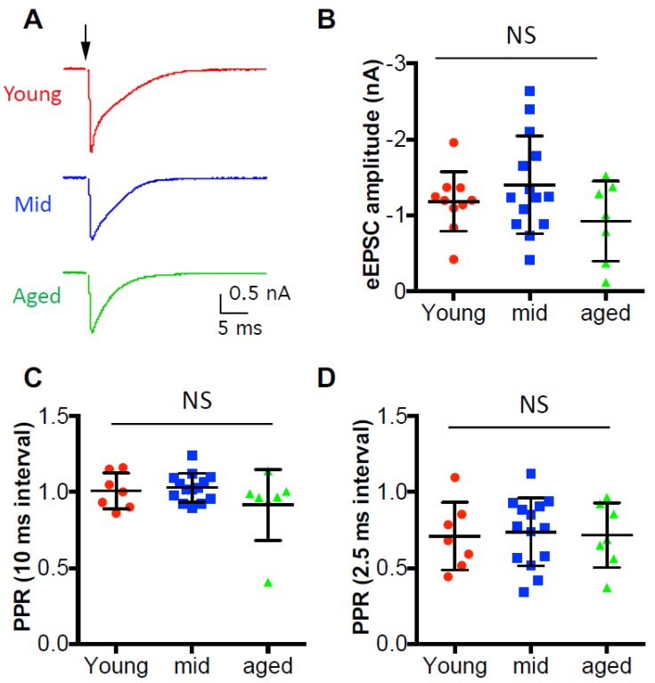
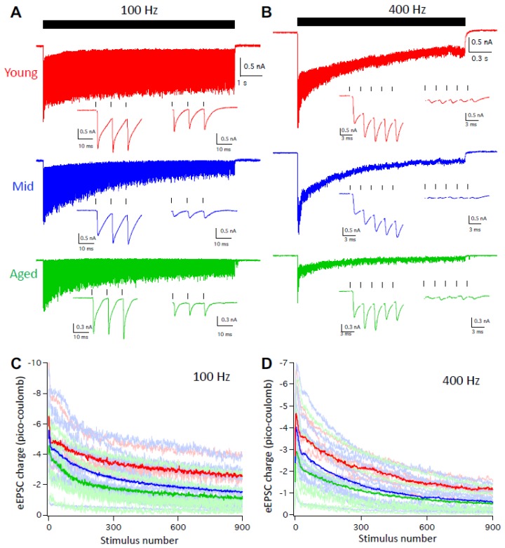
Age-related hearing loss (ARHL) is associated with weakened inhibition in the central auditory nervous system including the cochlear nucleus. One of the main inhibitory neurons of the cochlear nucleus is the D-stellate neuron, which provides extensive glycinergic inhibition within the local neural network. It remains unclear how physiological activities of D-stellate neurons change during ARHL and what are the underlying mechanisms. Using in vitro whole-cell patch clamp technique, we studied the intrinsic membrane properties of D-stellate neurons, the changes of their firing properties, and the underlying mechanisms in CBA/CaJ mice at the ages of 3-4 months (young), 17-19 months (middle age), and 27-33 months (aged). We found that the intrinsic membrane properties of D-stellate neurons were unchanged among these three age groups. However, these neurons showed decreased firing rate with age in response to sustained auditory nerve stimulation. Further investigation showed that auditory nerve-evoked excitatory postsynaptic currents (EPSCs) were significantly reduced in strength with age. These findings suggest that D-stellate neurons receive weakened synaptic inputs from the auditory nerve and decreased sound driven activity with age, which are expected to reduce the overall inhibition and enhance the central gain in the cochlear nucleus during ARHL.

Keywords: D-stellate; EPSC; age-related hearing loss; auditory nerve; firing rate; intrinsic property; synaptic transmission.


Conflict of interest statement

The authors declare no conflict of interest.

Figures

Figure 1
Figure 1
Auditory brainstem responses reveal age-related hearing loss in CBA/CaJ mice. (A) Representative auditory brainstem response (ABR) waveforms from mice in three age groups in response to clicks. Dots mark the traces at hearing threshold. (B) Summary ABR thresholds from all mice. Kruskal–Wallis test: * p < 0.05; *** p < 0.001; **** p < 0.0001. Individual data points are shown along with mean ± S.D. for each group.
Figure 2
Figure 2
D-stellate neurons show no change in intrinsic membrane properties with age. (AC) Example membrane responses of D-stellate neurons to current injections from three age groups. Numbers mark the resting membrane potentials in mV. Bottom panels: current pulse injections. (DL) Summary plots of all D-stellate neurons in resting membrane potential (D), membrane input resistance (E), membrane time constant (F), threshold current injection to trigger spikes (G), depolarization sag steady state versus peak ratio (H), depolarization sag decay time constant (I), spike amplitude (J), spike halfwidth (K) and spike threshold voltage (L). Kruskal–Wallis test (non-parametric one-way ANOVA) showed no significant difference in all measurements in (DL) (p > 0.05).
Figure 3
Figure 3
D-stellate neurons decrease in firing rate to auditory nerve inputs during aging. (A) Left: example responses of D-stellate neurons to auditory nerve inputs at 100 Hz. Ticks on top mark the timing of 900 stimuli at the auditory nerve. Note the decreased firing of spikes in middle and aged cells. Right: PSTH plots of the responses from the cells in left panels. (B) Same as in (A) except that the stimulus trains were at 400 Hz. (C) Normalized firing rate to 100 Hz stimulus trains in all D-stellate neurons from three age groups. Thin lines: individual neurons; thick lines: average firing rate in three age groups. (D) Normalized firing rates to 400 Hz stimulus trains.
Figure 4
Figure 4
D-stellate neurons show decreased firing rate with age but no change in temporal coding. (A) Firing rates in all D-stellate neurons to 100 Hz stimulus trains at the auditory nerve. (B) Firing rates in all D-stellate neurons to 400 Hz stimulus trains at the auditory nerve. (C) Calculated vector strength of D-stellate spikes evoked by 100 Hz stimulus trains at the auditory nerve. (D) Calculated vector strength of D-stellate spikes evoked by 400 Hz stimulus trains at the auditory nerve. Kruskal–Wallis test (non-parametric one-way ANOVA): ** p < 0.01; * p < 0.05; NS: p > 0.05.
Figure 5
Figure 5
Synaptic inputs to D-stellate neurons under quiescence show no change during aging. (A) Single stimulation at the auditory nerve evoked excitatory postsynaptic current (EPSCs) in three example D-stellate neurons from three age groups. Arrow: stimulus onset. (B) Summary plot of the EPSC amplitude in all three groups. (C) Paired pulse ratio (PPR) of EPSC amplitudes at 10 ms interval. (D): Paired pulse ratio of EPSC amplitude at 2.5 ms interval. NS: not significant (Kruskal–Wallis test: p > 0.05).
Figure 6
Figure 6
D-stellate neurons receive weakened synaptic inputs at high rate auditory nerve stimulation during aging. (A) Auditory nerve evoked EPSC trains at 100 Hz in D-stellate neurons from three age groups. Ticks on top mark the timing of 900 stimulus train. Two insets show evoked EPSCs by the first and last three stimuli of the train, respectively. (B) Auditory nerve-evoked EPSC trains at 400 Hz in the same D-stellate neurons as in (A). Traces in (A) and (B) are averages of 5 trials. (C and D) summary plots of EPSC charge transfer in D-stellate neurons at 100 (C) and 400 Hz (D). Thin lines are individual neurons; thick lines are the averages of all neurons in three age groups (red: young; blue: middle age; green: aged).

References

    1. Cant N.B., Benson C.G. Parallel auditory pathways: Projection patterns of the different neuronal populations in the dorsal and ventral cochlear nuclei. Brain. Res. Bull. 2003;60:457–474. doi: 10.1016/S0361-9230(03)00050-9. - DOI - PubMed
    1. Xie R., Manis P.B. Target-specific IPSC kinetics promote temporal processing in auditory parallel pathways. J. Neurosci. 2013;33:1598–1614. doi: 10.1523/JNEUROSCI.2541-12.2013. - DOI - PMC - PubMed
    1. Caspary D.M., Backoff P.M., Finlayson P.G., Palombi P.S. Inhibitory inputs modulate discharge rate within frequency receptive fields of anteroventral cochlear nucleus neurons. J. Neurophysiol. 1994;72:2124–2133. doi: 10.1152/jn.1994.72.5.2124. - DOI - PubMed
    1. Altschuler R.A., Betz H., Parakkal M.H., Reeks K.A., Wenthold R.J. Identification of glycinergic synapses in the cochlear nucleus through immunocytochemical localization of the postsynaptic receptor. Brain Res. 1986;369:316–320. doi: 10.1016/0006-8993(86)90542-1. - DOI - PubMed
    1. Campagnola L., Manis P.B. A map of functional synaptic connectivity in the mouse anteroventral cochlear nucleus. J. Neurosci. 2014;34:2214–2230. doi: 10.1523/JNEUROSCI.4669-13.2014. - DOI - PMC - PubMed

LinkOut - more resources