. 2019 Oct 31;8(11):1363.
doi: 10.3390/cells8111363.

Laquinimod Supports Remyelination in Non-Supportive Environments

Affiliations

Laquinimod Supports Remyelination in Non-Supportive Environments

Stella Nyamoya et al. Cells. .

Abstract

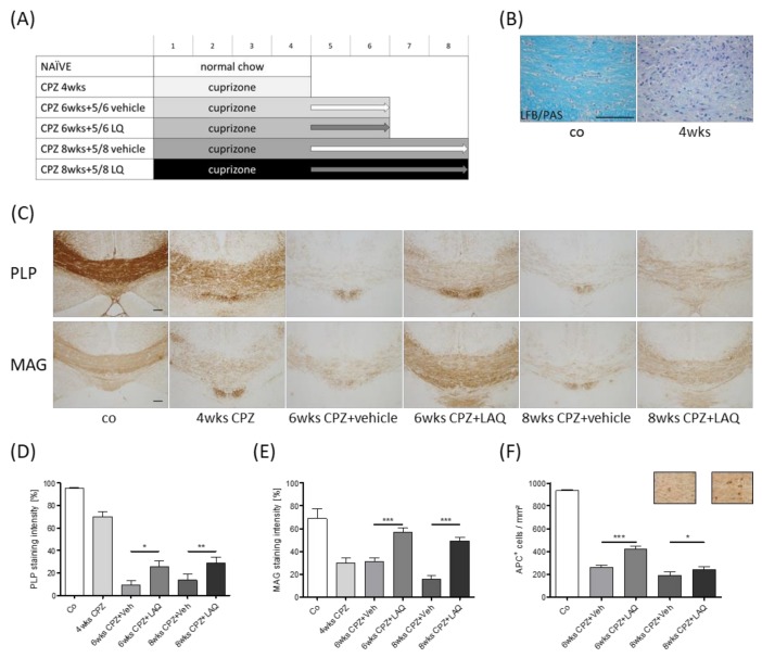
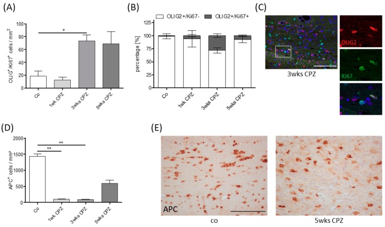
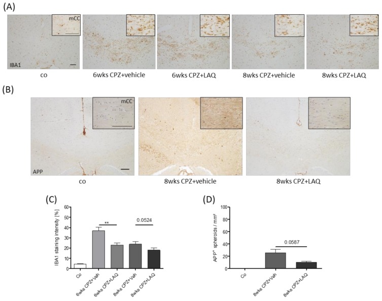
Inflammatory demyelination, which is a characteristic of multiple sclerosis lesions, leads to acute functional deficits and, in the long term, to progressive axonal degeneration. While remyelination is believed to protect axons, the endogenous-regenerative processes are often incomplete or even completely fail in many multiple sclerosis patients. Although it is currently unknown why remyelination fails, recurrent demyelination of previously demyelinated white matter areas is one contributing factor. In this study, we investigated whether laquinimod, which has demonstrated protective effects in active multiple sclerosis patients, protects against recurrent demyelination. To address this, male mice were intoxicated with cuprizone for up to eight weeks and treated with either a vehicle solution or laquinimod at the beginning of week 5, where remyelination was ongoing. The brains were harvested and analyzed by immunohistochemistry. At the time-point of laquinimod treatment initiation, oligodendrocyte progenitor cells proliferated and maturated despite ongoing demyelination activity. In the following weeks, myelination recovered in the laquinimod- but not vehicle-treated mice, despite continued cuprizone intoxication. Myelin recovery was paralleled by less severe microgliosis and acute axonal injury. In this study, we were able to demonstrate that laquinimod, which has previously been shown to protect against cuprizone-induced oligodendrocyte degeneration, exerts protective effects during oligodendrocyte progenitor differentiation as well. By this mechanism, laquinimod allows remyelination in non-supportive environments. These results should encourage further clinical studies in progressive multiple sclerosis patients.

Keywords: cuprizone; laquinimod; multiple sclerosis; neurodegeneration; remyelination.


Conflict of interest statement

The authors declare no conflict of interest

Figures

Figure 1
Figure 1
Influence of laquinimod on remyelination in the non-supportive environment. (A) Schematic depiction of the experimental setup. Numbers indicate the duration of the experiment in weeks. The control group is colored in white, cuprizone (CPZ) intoxication groups are colored in grayscale and black. The arrows indicate treatment with either the vehicle (Veh, light gray) or laquinimod (LAQ, dark gray) solutions. (B) Representative image of Luxol fast blue / periodic acid–Schiff (LFB/PAS) staining of a control animal and an animal intoxicated with cuprizone for 4 weeks. (C) Representative images of anti-PLP and anti-MAG stained sections of the midline corpus callosum. Densitometric analysis of (D) anti-PLP and (E) anti-MAG staining intensity (repetitive Mann–Whitney test, as indicated). (F) Quantification of APC+ cell numbers (repetitive Mann Whitney test as indicated). Representative images of anti-APC immunohistochemical stained sections of the medial corpus callosum of 8 weeks cuprizone plus vehicle (left) or laquinimod (right) treatment groups. Scale bar: 100 μm. The following symbols were used to indicate the level of significance: * p < 0.05, ** p < 0.005, and *** p < 0.001.
Figure 2
Figure 2
Oligodendrocyte pathology. (A) Quantity of OLIG2+ and Ki67+ single and double positive cells during the course of cuprizone-induced demyelination (Kruskal–Wallis test followed by Dunn’s multiple comparison test). (B) Percentage of single and double positive cells in relation to the entire OLIG2+ cell population. (C) Representative image of OLIG2+ and Ki67+ double positive cells. (D) Quantification of APC+ cell numbers (Kruskal-Wallis test followed by Dunn’s multiple comparison test). (E) Representative images of anti-APC stained sections in the medial corpus callosum. Scale bar: 100 μm. The following symbols were used to indicate the level of significance: * p < 0.05, ** p < 0.005.
Figure 3
Figure 3
Microglia/monocyte accumulation and axonal damage. (A) Accumulation of microglia/monocytes visualized by anti-IBA1 immunohistochemistry. Inserts show the medial corpus callosum in higher magnification. (B) Axonal injury visualized by anti-APP immunohistochemistry. (C) Densitometric analysis of anti-IBA1 stains (Mann–Whitney test). (D) APP+ spheroid density quantification (Mann–Whitney test). Scale bar: 100μm. The following symbols were used to indicate the level of significance: ** p < 0.005.

References

    1. Haider L., Simeonidou C., Steinberger G., Hametner S., Grigoriadis N., Deretzi G., Frischer J.M. Multiple sclerosis deep grey matter: The relation between demyelination, neurodegeneration, inflammation and iron. J. Neurol Neurosurg Psychiatry. 2014;85:1386–1395. doi: 10.1136/jnnp-2014-307712. - DOI - PMC - PubMed
    1. Ferguson B., Matyszak M.K., Esiri M.M., Perry V.H. Axonal damage in acute multiple sclerosis lesions. Brain. 1997;120:393–399. doi: 10.1093/brain/120.3.393. - DOI - PubMed
    1. Kuhlmann T., Lingfeld G., Bitsch A., Schuchardt J., Bruck W. Acute axonal damage in multiple sclerosis is most extensive in early disease stages and decreases over time. Brain. 2002;125:2202–2212. doi: 10.1093/brain/awf235. - DOI - PubMed
    1. Bauer J., Rauschka H., Lassmann H. Inflammation in the nervous system: The human perspective. Glia. 2001;36:235–243. doi: 10.1002/glia.1112. - DOI - PubMed
    1. Barnett M.H., Prineas J.W. Relapsing and remitting multiple sclerosis: Pathology of the newly forming lesion. Ann. Neurol. 2004;55:458–468. doi: 10.1002/ana.20016. - DOI - PubMed

Publication types

MeSH terms

LinkOut - more resources