

Roles for Autophagy in Esophageal Carcinogenesis: Implications for Improving Patient Outcomes
- PMID: 31683722
- PMCID: PMC6895837
- DOI: 10.3390/cancers11111697
Roles for Autophagy in Esophageal Carcinogenesis: Implications for Improving Patient Outcomes
Abstract
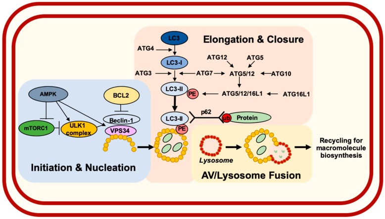
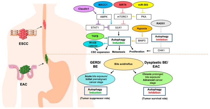
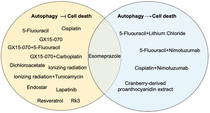
Esophageal cancer is among the most aggressive forms of human malignancy with five-year survival rates of <20%. Autophagy is an evolutionarily conserved catabolic process that degrades and recycles damaged organelles and misfolded proteins to maintain cellular homeostasis. While alterations in autophagy have been associated with carcinogenesis across tissues, cell type- and context-dependent roles for autophagy have been reported. Herein, we review the current knowledge related to autophagy in esophageal squamous cell carcinoma (ESCC) and esophageal adenocarcinoma (EAC), the two most common subtypes of esophageal malignancy. We explore roles for autophagy in the development and progression of ESCC and EAC. We then continue to discuss molecular markers of autophagy as they relate to esophageal patient outcomes. Finally, we summarize current literature examining roles for autophagy in ESCC and EAC response to therapy and discuss considerations for the potential use of autophagy inhibitors as experimental therapeutics that may improve patient outcomes in esophageal cancer.
Keywords: autophagy; esophageal adenocarcinoma; esophageal cancer; esophageal squamous cell carcinoma; esophagus.
Conflict of interest statement
The authors declare no conflict of interest.
Figures
Similar articles
-
Autophagy as a cytoprotective mechanism in esophageal squamous cell carcinoma.Curr Opin Pharmacol. 2018 Aug;41:12-19. doi: 10.1016/j.coph.2018.04.003. Epub 2018 Apr 17. Curr Opin Pharmacol. 2018. PMID: 29677645 Free PMC article. Review.
-
Comparative genomic analysis of esophageal squamous cell carcinoma and adenocarcinoma: New opportunities towards molecularly targeted therapy.Acta Pharm Sin B. 2022 Mar;12(3):1054-1067. doi: 10.1016/j.apsb.2021.09.028. Epub 2021 Sep 30. Acta Pharm Sin B. 2022. PMID: 35530133 Free PMC article. Review.
-
Comprehensive Genomic Profiling of Advanced Esophageal Squamous Cell Carcinomas and Esophageal Adenocarcinomas Reveals Similarities and Differences.Oncologist. 2015 Oct;20(10):1132-9. doi: 10.1634/theoncologist.2015-0156. Epub 2015 Sep 2. Oncologist. 2015. PMID: 26336083 Free PMC article.
-
Autophagy and its current relevance to the diagnosis and clinical management of esophageal diseases.Ann N Y Acad Sci. 2016 Oct;1381(1):113-121. doi: 10.1111/nyas.13190. Epub 2016 Aug 15. Ann N Y Acad Sci. 2016. PMID: 27526024 Review.
-
Autophagy and Apoptosis Play Opposing Roles in Overall Survival of Esophageal Squamous Cell Carcinoma.Pathol Oncol Res. 2016 Oct;22(4):699-705. doi: 10.1007/s12253-016-0051-z. Epub 2016 Mar 15. Pathol Oncol Res. 2016. PMID: 26980476
Cited by
-
Autophagy: Shedding Light on the Mechanisms and Multifaceted Roles in Cancers.Biomolecules. 2025 Jun 22;15(7):915. doi: 10.3390/biom15070915. Biomolecules. 2025. PMID: 40723786 Free PMC article. Review.
-
Construction and Evaluation of a Risk Score Model for Autophagy-Related Genes in Esophageal Adenocarcinoma.Med Sci Monit. 2021 Jan 29;27:e927850. doi: 10.12659/MSM.927850. Med Sci Monit. 2021. PMID: 33510126 Free PMC article.
-
Establishment and Analysis of a Prognostic Model of Autophagy-Related lncRNAs in ESCA.Biomed Res Int. 2022 Jul 26;2022:9265088. doi: 10.1155/2022/9265088. eCollection 2022. Biomed Res Int. 2022. PMID: 35928921 Free PMC article.
-
Autophagy: New Insights into Its Roles in Cancer Progression and Drug Resistance.Cancers (Basel). 2020 Oct 16;12(10):3005. doi: 10.3390/cancers12103005. Cancers (Basel). 2020. PMID: 33081217 Free PMC article.
-
Defining the Role of GLI/Hedgehog Signaling in Chemoresistance: Implications in Therapeutic Approaches.Cancers (Basel). 2021 Sep 23;13(19):4746. doi: 10.3390/cancers13194746. Cancers (Basel). 2021. PMID: 34638233 Free PMC article. Review.
References
Publication types
Grants and funding
LinkOut - more resources
Full Text Sources
