. 2019 Nov 1;10(11):882.
doi: 10.3390/genes10110882.

The BRCA1 c.4096+3A>G Variant Displays Classical Characteristics of Pathogenic BRCA1 Mutations in Hereditary Breast and Ovarian Cancers, But Still Allows Homozygous Viability

Affiliations

The BRCA1 c.4096+3A>G Variant Displays Classical Characteristics of Pathogenic BRCA1 Mutations in Hereditary Breast and Ovarian Cancers, But Still Allows Homozygous Viability

Adalgeir Arason et al. Genes (Basel). .

Abstract

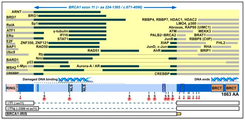
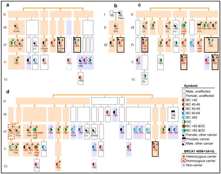
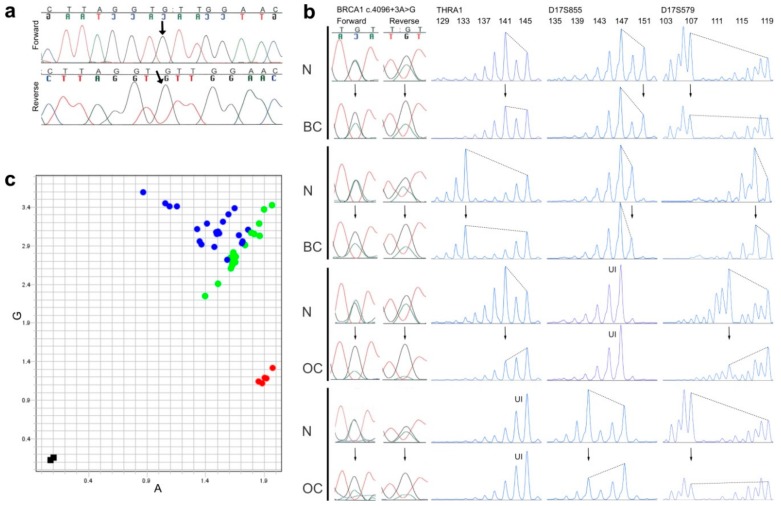
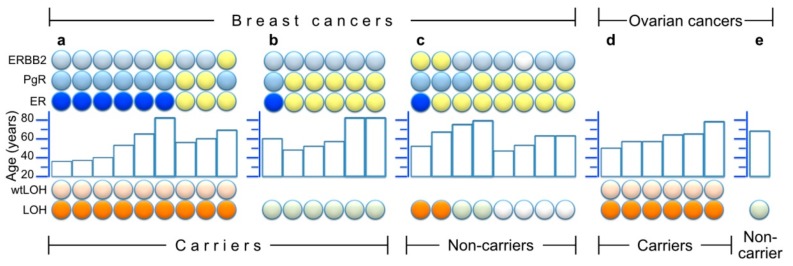
Mutations in BRCA1 result in predisposal to breast and ovarian cancers, but many variants exist with unknown clinical significance (VUS). One is BRCA1 c.4096+3A>G, which affects production of the full-length BRCA1 transcript, while augmenting transcripts lacking most or all of exon 11. Nonetheless, homozygosity of this variant has been reported in a healthy woman. We saw this variant cosegregate with breast and ovarian cancer in several family branches of four Icelandic pedigrees, with instances of phenocopies and a homozygous woman with lung cancer. We found eight heterozygous carriers (0.44%) in 1820 unselected breast cancer cases, and three (0.15%) in 1968 controls (p = 0.13). Seeking conclusive evidence, we studied tumors from carriers in the pedigrees for wild-type-loss of heterozygosity (wtLOH) and BRCA1-characteristic prevalence of estrogen receptor (ER) negativity. Of 15 breast and six ovarian tumors, wtLOH occurred in nine breast and all six ovarian tumours, and six of the nine breast tumors with wtLOH were ER-negative. These data accord with a pathogenic BRCA1-mutation. Our findings add to the current knowledge of BRCA1, and the role of its exon 11 in cancer pathogenicity, and will be of use in clinical genetic counselling.

Keywords: BRCA1; Knudson's two-hit model; LOH; VUS; breast cancer; cancer risk; homozygous lethality; ovarian cancer; tumorigenesis.


Conflict of interest statement

The authors declare no conflict of interest.

Figures

Figure 1
Figure 1
Domain map of the full-length BRCA1 protein (1863 amino acids), emphasizing its middle exon-11-derived region (light blue; bulged if included in Δ11q, but not Δ11 isoforms), which contains two nuclear localization signals (NLS), a coiled-coil domain (CCD), eight serine/threonine phosphorylation spots (pinned S or T on an encircled P), and binding positions for proteins (yellow area, black bars) and for damaged DNA. For comparison, indicated to the left and right of exon 11, are additional protein binding positions (yellow area, grey bars), a position of binding to DNA ends, further serine phosphorylation spots (smaller) and the following domains: RING (which binds, e.g., BARD1), NES (nuclear export signal), BRCT phosphopeptide recognition domains, and a CCD that mediates interaction with PALB2, which bridges to BRCA2. Below, in boxes, are the parts contained in smaller isoforms (connected by broken lines for skipped parts), with the intron-11-derived C-tail of BRCA1-IRIS (bottom) indicated by a small yellow/orange box (not present in full-length BRCA1 or other isoforms).
Figure 2
Figure 2
Pedigrees (ad) of eight probands (arrows in boxes with black borders), showing cancer-affected individuals and their carrier status, with regard to the BRCA1 c.4096+3A>G variant, but withholding details on other pedigree members and exact pedigree structures, in order to avoid recognition. Light orange background is used to highlight segregation of the variant, and light violet background highlights cancer-affected non-carriers. Individuals with a white background were not available for genotyping. See explanation of symbols in the lower right corner of the figure (BC = breast cancer, OC = ovarian cancer). Information about approximate age (in years) at diagnosis of cancer is shown below symbols, in accordance with the symbol’s indication of cancer type, plus further details, in case of contralateral breast cancer (cont), fallopian tubes (Fall), endodermal sinus (Es) or cancers other than BC or OC (Bra for brain, Col for colorectal, Emet for endometrial, Esop for esophageal, Glb for gallbladder, Lu for lung, Pro for prostatic, Stom for stomach, Thyr for thyroid, Uter for uterine). Letters in bold red italic denote that the tumor in question was included in analyses of LOH (loss of heterozygosity) and/or histology.
Figure 3
Figure 3
Genotyping and LOH results. (a) Sanger sequencing (forward and reverse electropherograms) of the BRCA1 c.4096+3A>G locus in a homozygous carrier (see Figure 2d, far left in generation IV), the variant signal is indicated by an arrow; (b) Examples of pairs of normal (N) and breast (BC) or ovarian cancer tissue (OC) from the same individual, compared for possible loss of heterozygosity in tumor tissue (arrows pointing to altered signal heights in tumors), as analyzed by Sanger sequencing of the BRCA1 c.4096+3A>G locus (left part, both forward and reverse electropherograms shown) and by PCR-fragment analyses of three microsatellite markers within and flanking the BRCA1 gene (THRA1, D17S855 and D17S579, with allele sizes in base pairs shown on the top, and UI denoting homozygous genotypes, which were uninformative about LOH); (c) An allelic discrimination plot from a BRCA1 c.4096+3A>G SNP assay of 21 NT-pairs (pairs of matched normal- and tumor-tissue-derived DNA from the same individual) from individuals who were heterozygous for the BRCA1 c.4096+3A>G variant. Instead of conventional automatic coloring of genotypes provided by the manufacturer’s software, the dots in this graph were manually colored green if they derived from normal tissue and blue if they derived from tumor tissue. Five samples from non-carriers (homozygous for the A-allele) are shown in red, and two negative controls (water) in black.
Figure 4
Figure 4
Summary of LOH results and/or histological staining of estrogen-, progesterone- and HER2/Erb-B2 receptors (ER, PgR and ERBB2, respectively) in 30 tumors. Within each partition of the figure, the samples are ordered from left to right by LOH results (bottom symbols: orange for LOH and light orange above that, denoting wtLOH, light green for samples without LOH, and white for unavailable LOH information), and then by ER results (ER line of symbols: blue for ER negative, yellow for ER positive) and finally by age (middle histogram) and other receptor results (top two lines of symbols: light blue = negative, yellow = positive and white = unavailable). (a) Nine breast cancers from heterozygous carriers who had wtLOH in their tumors; (b) Six breast cancers from heterozygous carriers without LOH; (c) Eight breast cancers from non-carriers among close (1st and 2nd degree) relatives of genotyped or obligate carriers (four of which were available for LOH analysis of microsatellite markers); (d) Six ovarian cancers from carriers; (e) One ovarian cancer from a non-carrier.

References

    1. Lu Y., Ek W.E., Whiteman D., Vaughan T.L., Spurdle A.B., Easton D.F., Pharoah P.D., Thompson D.J., Dunning A.M., Hayward N.K., et al. Most common ‘sporadic’ cancers have a significant germline genetic component. Hum. Mol. Genet. 2014;23:6112–6118. doi: 10.1093/hmg/ddu312. - DOI - PMC - PubMed
    1. Garber J.E., Offit K. Hereditary cancer predisposition syndromes. J. Clin. Oncol. 2005;23:276–292. doi: 10.1200/JCO.2005.10.042. - DOI - PubMed
    1. Miki Y., Swensen J., Shattuck-Eidens D., Futreal P.A., Harshman K., Tavtigian S., Liu Q., Cochran C., Bennett L.M., Ding W., et al. A strong candidate for the breast and ovarian cancer susceptibility gene BRCA1. Science. 1994;266:66–71. doi: 10.1126/science.7545954. - DOI - PubMed
    1. Wooster R., Bignell G., Lancaster J., Swift S., Seal S., Mangion J., Collins N., Gregory S., Gumbs C., Micklem G., et al. Identification of the breast cancer susceptibility gene BRCA2. Nature. 1995;378:789–792. doi: 10.1038/378789a0. - DOI - PubMed
    1. Melchor L., Benitez J. The complex genetic landscape of familial breast cancer. Hum. Genet. 2013;132:845–863. doi: 10.1007/s00439-013-1299-y. - DOI - PubMed

Publication types