. 2019 Nov 2;8(11):541.
doi: 10.3390/foods8110541.

Optimization of a Microplate Assay for Generating Listeria Monocytogenes, E. Coli O157:H7, and Salmonella Biofilms and Enzymatic Recovery for Enumeration

Affiliations

Optimization of a Microplate Assay for Generating Listeria Monocytogenes, E. Coli O157:H7, and Salmonella Biofilms and Enzymatic Recovery for Enumeration

Manish Aryal et al. Foods. .

Abstract

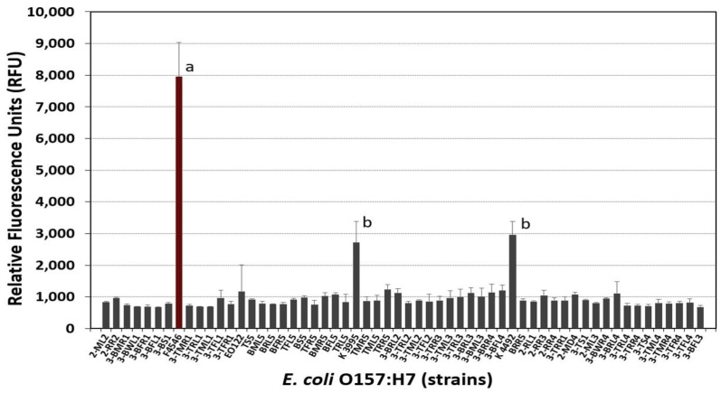
Biofilms enable the persistence of pathogens in food processing environments. Sanitizing agents are needed that are effective against pathogens entrapped in biofilms that are more difficult to inactivate than planktonic cells that are displaced and found on equipment surfaces. We examined conditions to develop, analyze, and enumerate the enhanced biofilms of three different foodborne pathogens assisted by fluorescence adherence assay and enzymatic detachment. We compared three different isomeric forms of fluorescent substrates that are readily taken up by bacterial cells based on carboxy-fluorescein diacetate (5-CFDA, 5,6-CFDA, 5,6-CFDA, SE). Biofilm-forming strains of Escherichia coli O157:H7 F4546 and Salmonella Montevideo FSIS 051 were identified using a microplate fluorescence assay defined previously for L. monocytogenes. Adherence levels were determined by differences in relative fluorescence units (RFU) as well as recovered bacterial cells. Multiple hydrolytic enzymes were examined for each representative pathogen for the most suitable enzyme for detachment and enumeration to confirm adherence data obtained by fluorescence assay. Cultures were grown overnight in microplates, incubated, washed and replenished with fresh sterile growth medium; this cycle was repeated for seven consecutive days to enrich for robust biofilms. Treatments were performed in triplicate and compared by one-way analysis of variance (ANOVA) to determine significant differences (p < 0.05).

Keywords: E. coli O157:H7; Listeria monocytogenes; Salmonella; biofilm; carboxyfluorescein diacetate; enzymes; microplate assay.


Conflict of interest statement

The authors declare no conflict of interest.

Figures

Figure 1
Figure 1
(A) Comparison of fluorescence signals obtained using L. monocytogenes 99-38 in microplate fluorescence assay with 5,6-CFDA, 5,6-CFDA, SE, or 5-CFDA. Data are presented as the mean of triplicate replications and error bars represent the standard deviation from the mean. Means with different letters are significantly different as determined by one-way ANOVA using the Holm-Sidak test for pairwise multiple comparisons to determine significant differences (p < 0.05). (B) Two-fold dilutions of planktonic cells incubated with 5,6-CFDA compared to cells without 5,6-CFDA and examined for fluorescence signals (Ex/Em: 485/535 nm). Error bars are the standard deviation of the means of triplicate replications.
Figure 2
Figure 2
Comparison of adherence of (A) Listeria monocytogenes and (B) Salmonella by microplate fluorescence assay with 5,6-CFDA. Data are presented as the mean of triplicate replications and error bars represent the standard deviation from the mean. Means with different letters are significantly different as determined by one-way ANOVA using the Holm-Sidak test for pairwise multiple comparisons to determine significant differences (p < 0.05); means with the same letter are not significantly different (p > 0.05).
Figure 3
Figure 3
Comparison of adherence of various strains of E. coli O157:H7 by microplate fluorescence assay with 5,6-CFDA. Data are presented as the mean of triplicate replications and error bars represent the standard deviation from the mean. Means with different letters are significantly different as determined by one-way ANOVA using the Holm-Sidak test for pairwise multiple comparisons to determine significant differences (p < 0.05); means with the same letter are not significantly different (p > 0.05).
Figure 4
Figure 4
Enumeration of viable cells after multiple buffer washes with 0.05 M Tris buffer (pH 7.4) and after Bax protease treatment (after final wash) of L. monocytogenes 99-38 microplate biofilms. Data are presented as the mean of triplicate replications and error bars represent the standard deviation from the mean. Means with different letters are significantly different as determined by one-way ANOVA using the Holm-Sidak test for pairwise multiple comparisons to determine significant differences (p < 0.05); means with the same letter are not significantly different (p > 0.05).
Figure 5
Figure 5
Comparison of enumeration and fluorescence data with L. monocytogenes 99-38, Salmonella Montevideo FSIS 051, and E. coli F4546, before and after enzyme treatment of microplate biofilms. (A) Cell enumeration after 3rd round wash buffer followed by Bax protease release of adhered cells from microplates. (B) Fluorescence of biofilms with 5,6-CFDA before and after Bax protease treatment to release bacterial cells. Data are presented as the mean of quadruple replications and error bars represent the standard deviation from the mean. Significant differences are between treatments with the same strain. Means with different letters are significantly different as determined by one-way ANOVA using the Holm-Sidak test for pairwise multiple comparisons to determine significant differences (p < 0.05).
Figure 6
Figure 6
Enumeration of L. monocytogenes 99-38 biofilm levels over time after repeated incubation in microplates. Planktonic cells were removed daily, washed with buffer, and replaced with fresh sterile media. Attached cells were enumerated by detachment with pronase E and represented as the means of triplicate replications; error bars represent the standard deviation of the means. Means with different letters are significantly different as determined by one-way ANOVA using the Holm-Sidak test for pairwise multiple comparisons to determine significant differences (p < 0.05); means with the same letter are not significantly different (p > 0.05).
Figure 7
Figure 7
Recovery and enumeration of L. monocytogenes 99-38, E. coli O157:H7 F4546, and Salmonella Montevideo FSIS 051 biofilms after treatment with various enzymes (Bax protease, cellulase, pronase E, papain, trypsin, or lipase). Data bars represent the means of triplicate replications and error bars represent standard deviation of the means. Means with different letters are significantly different as determined by one-way ANOVA using the Holm-Sidak test for pairwise multiple comparisons within the same organism to determine significant differences (p < 0.05); means with the same letter are not significantly different (p > 0.05).

Similar articles

Cited by

References

    1. Zhao X., Zhao F., Wang J., Zhong N. Biofilm formation and control strategies of foodborne pathogens: Food safety perspectives. RSC Adv. 2017;7:36670–36683. doi: 10.1039/C7RA02497E. - DOI
    1. Achinas S.C.N., Euverink G.J.W. A brief recap of microbial adhesion and biofilms. Appl. Sci. 2019;9:2801. doi: 10.3390/app9142801. - DOI
    1. Stoodley P., Sauer K., Davies D.G., Costerton J.W. Biofilms as complex differentiated communities. Annu. Rev. Microbiol. 2002;56:187–209. doi: 10.1146/annurev.micro.56.012302.160705. - DOI - PubMed
    1. Hinsa S.M., Espinosa-Urgel M., Ramos J.L., O’Toole G.A. Transition from reversible to irreversible attachment during biofilm formation by Pseudomonas fluorescens WCS365 requires an ABC transporter and a large secreted protein. Mol. Microbiol. 2003;49:905–918. doi: 10.1046/j.1365-2958.2003.03615.x. - DOI - PubMed
    1. Wagner S., Hauck D., Hoffmann M., Sommer R., Joachim I., Müller R., Imberty A., Varrot A., Titz A. Covalent lectin inhibition and application in bacterial biofilm imaging. Angew. Chem. Int. Ed. 2017;56:16559–16564. doi: 10.1002/anie.201709368. - DOI - PMC - PubMed

LinkOut - more resources