Review
. 2019 Nov 3;8(11):1380.
doi: 10.3390/cells8111380.

Wnt Signaling in the Regulation of Immune Cell and Cancer Therapeutics

Affiliations
Review

Wnt Signaling in the Regulation of Immune Cell and Cancer Therapeutics

Muhammad Haseeb et al. Cells. .

Abstract

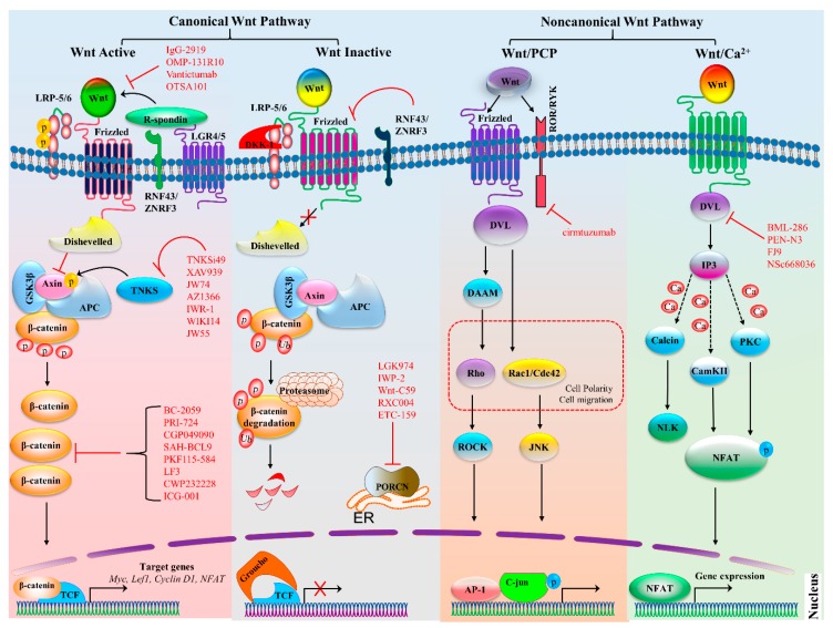
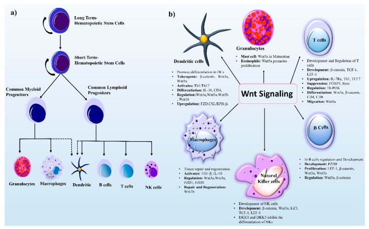
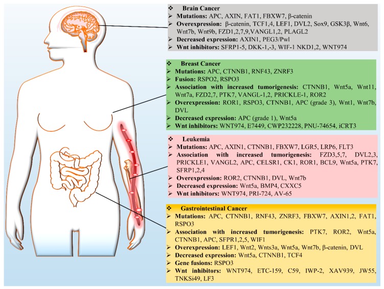
Wnt signaling is one of the important pathways to play a major role in various biological processes, such as embryonic stem-cell development, tissue regeneration, cell differentiation, and immune cell regulation. Recent studies suggest that Wnt signaling performs an essential function in immune cell modulation and counteracts various disorders. Nonetheless, the emerging role and mechanism of action of this signaling cascade in immune cell regulation, as well as its involvement in various cancers, remain debatable. The Wnt signaling in immune cells is very diverse, e.g., the tolerogenic role of dendritic cells, the development of natural killer cells, thymopoiesis of T cells, B-cell-driven initiation of T-cells, and macrophage actions in tissue repair, regeneration, and fibrosis. The purpose of this review is to highlight the current therapeutic targets in (and the prospects of) Wnt signaling, as well as the potential suitability of available modulators for the development of cancer immunotherapies. Although there are several Wnt inhibitors relevant to cancer, it would be worthwhile to extend this approach to immune cells.

Keywords: Wnt signaling; cancer; immune cell regulation; inhibitor; therapeutic target.


Conflict of interest statement

The authors declare that there are no competing interests.

Figures

Figure 1
Figure 1
Canonical and noncanonical Wnt signaling pathways. The canonical Wnt pathway is activated by the binding of a Wnt ligands to a frizzled (FZD) family receptor and co-receptor LRP5 or LRP6, which recruits disheveled (Dvl), consequently inactivating the destruction complex composed of APC, GSK3β, and axin. This inactivation prevents β-catenin from proteasomal degradation and allows for the accumulation of β-catenin, which then enters the nucleus. There, it binds to transcription factor TCF or LEF and initiates the transcription of target genes. Tankyrases (TNKSs) also promote signaling by targeting axin for degradation. Moreover, when R-spondin binds to LGR4 or LGR5, RNF43, and ZNRF3, it is not capable of targeting FZD family receptors for degradation and enhances Wnt signaling. There are various inhibitors of the Wnt signaling pathway, particularly those targeting Wnt ligands, Dvl, TNKS, β-catenin, and PORCN, which are highlighted in red; bars indicate the inhibitory effect. In the absence of Wnt ligands, the destruction complex becomes active and starts the proteasomal degradation of β-catenin. Proteins RNF43 and ZNRF3 also inhibit the binding of FZD and target it for degradation. The noncanonical Wnt–PCP pathway is triggered by Wnt ligands that increase the heterodimerization of a receptor-like tyrosine kinase (RYK) and tyrosine kinase–like orphan receptor (ROR). The binding to the receptor activates the Dvl protein and downstream signaling, for instance, DAAM activates GTPases Rho and ROCK, whereas the activation of c-Jun N-terminal kinase (JNK) by Rac is independent of DAAM. They collectively regulate cell polarity and migration and have also been implicated in cancer. The Wnt–Ca2+ pathway is activated by ligand Wnt, which raises the intracellular Ca2+ levels and generates inositol 1,4,5-triphosphate-3 (IP3). The Ca2+ levels increase and switch on downstream Ca2+-dependent enzymes such as calmodulin-dependent protein kinase (CaMKII), calcineurin, and protein kinase C (PKC). As a consequence, CaMKII and PKC phosphorylate nuclear factor of activated T cells (NFAT) and activate the expression of target genes. Protein symbols and abbreviations: APC, adenomatous polyposis coli protein; AP-1, activator protein 1; DKK1, dickkopf related protein 1; ER, endoplasmic reticulum; GSK3β, glycogen synthase kinase 3β; LEF1, lymphoid enhancer-binding factor 1; LRP, lipoprotein receptor-related protein; LGR4/5, Leucine-rich-repeat–containing G protein–coupled receptor 4 or 5; NLK, Nemo like kinase; RNF43, Ring finger protein 43; and ZNRF3, zinc ring finger 3.
Figure 2
Figure 2
HSC development and Wnt signaling components in immune cell regulation. (a) Differentiated blood cells are generated from self-renewing LT-HSCs, which are capable of differentiation and self-renewal. When LT-HSCs differentiate, they form ST-HSCs with a limited self-renewal capability. The ST-HSCs next produce the multipotent non self-renewing common myeloid linage (granulocytes and macrophages) and common lymphoid lineage (B cells, T cells, and NK cells). The dashed lines show partial progenitor connections. (b) Wnt signaling components play role in myeloid and lymphoid lineage cells. Wnt ligands (Wnt3a, Wnt5a, Wnt5b, and Wnt16) and receptors (FZD1 and FZD5) take part in the regulation of immune cells. The major role of Wnt signaling in B cells, T cells, and NK cells is development. By contrast, in macrophages, this signaling governs tissue repair and regeneration. Wnt signaling and its components perform different tasks in immune cells such as activation, proliferation, migration, tolerogenesis, and up- and down-regulation of genes as shown in text boxes. Protein symbols and abbreviations: CD4, cluster of differentiation 4; DCs, dendritic cells; DKK, dickkopf-related protein; FZD, frizzled; FOXP3, forkhead box P3; IL-10, interleukin 10; IL7Ra, interleukin 7 receptor α; LEF1, lymphoid enhancer-binding factor 1; LiC1, ligand-gated ion channel; NKs, natural Killer cells; RORC, related orphan receptor C; TH1, T helper 1 cell; TGFβ, transforming growth factor β; Th-POK, a zinc finger protein.
Figure 3
Figure 3
Wnt signaling in cancers. Deregulation of Wnt signaling components is involved in selected cancers (brain cancer, leukemia, breast cancer, and GCs) via a variety of mechanisms, including mutations, overexpression of Wnt proteins, decreased expression, gene fusion, and increased tumorigenesis; these data are summarized in the boxes. Protein symbols: BCL9, B-cell CLL or lymphoma 9; BMP4, bone morphogenetic protein 4; CELSR1, cadherin EGF LAG seven-pass G-type receptor 1; FBXW7, F-box and WD Repeat Domain -containing 7; FLT3, Fms-related tyrosine kinase 3; NKD1, naked cuticle homolog 1; PEG3/Pwl, paternally expressed 3; PLAG2, pleiomorphic adenoma-like protein 2; SFRP1, secreted frizzled-related protein 1; VANGL, Vang-like protein; WIF-1, Wnt-inhibitory factor 1.

References

    1. Barker N. Adult intestinal stem cells: Critical drivers of epithelial homeostasis and regeneration. Nat. Rev. Mol. Cell Biol. 2014;15:19–33. doi: 10.1038/nrm3721. - DOI - PubMed
    1. Clevers H., Nusse R. Wnt/beta-catenin signaling and disease. Cell. 2012;149:1192–1205. doi: 10.1016/j.cell.2012.05.012. - DOI - PubMed
    1. Staal F.J., Luis T.C., Tiemessen M.M. WNT signalling in the immune system: WNT is spreading its wings. Nat. Rev. Immunol. 2008;8:581–593. doi: 10.1038/nri2360. - DOI - PubMed
    1. Gieseck R.L., 3rd, Wilson M.S., Wynn T.A. Type 2 immunity in tissue repair and fibrosis. Nat. Rev. Immunol. 2018;18:62–76. doi: 10.1038/nri.2017.90. - DOI - PubMed
    1. Nusse R., Clevers H. Wnt/beta-Catenin Signaling, Disease, and Emerging Therapeutic Modalities. Cell. 2017;169:985–999. doi: 10.1016/j.cell.2017.05.016. - DOI - PubMed

Publication types

LinkOut - more resources