

The first case of gland inclusion in an intrapulmonary lymph node: a mimic of metastasis
- PMID: 31684955
- PMCID: PMC6829919
- DOI: 10.1186/s12957-019-1726-1
The first case of gland inclusion in an intrapulmonary lymph node: a mimic of metastasis
Abstract
Background: Lymph node inclusions are foci of ectopic tissue in lymph nodes, which were reported in different areas of the body. However, inclusions in the mediastinal lymph node are rare. Here, we report the first case of glandular inclusion within the parenchyma of the intrapulmonary lymph node in a patient with primary lung adenocarcinoma.
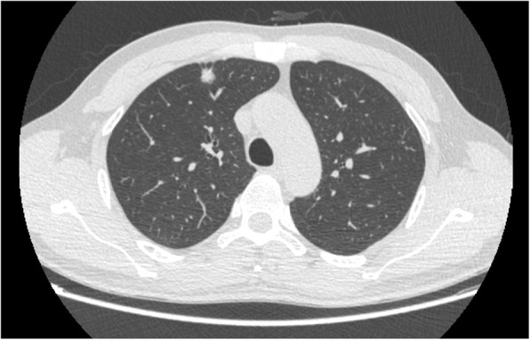
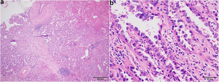
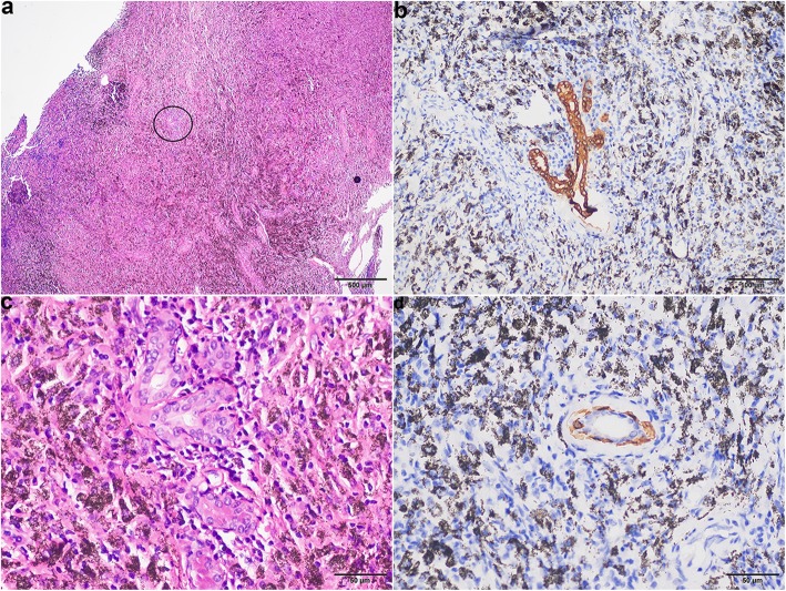
Case presentation: A computed tomography (CT) scan showed a solid pulmonary nodule in the right upper lobe in a 44-year-old man. After a fine needle aspiration biopsy diagnosis of adenocarcinoma, lobectomy and lymph dissection were performed. Histological sections of the lung demonstrated a papillary predominant adenocarcinoma and one intrapulmonary lymph node, which displayed glandular inclusion occupying the node parenchyma. The gland inclusion was very similar to metastasis, but was formed by two layers of epithelial cells, and the abluminal cells were positive for P63, P40, and CK5/6. The patient has remained alive without recurrence and metastasis at the last follow-up before publication.
Conclusions: It is very important to correctly diagnose a lymph node inclusion for proper clinical management.
Keywords: Epithelial inclusion; Lung adenocarcinoma; Lymph node; Metastasis; Tumour staging.
Conflict of interest statement
The authors declare that they have no competing interests.
Figures
Similar articles
-
Outcomes and predictive factors for pathological node-positive in radiographically pure-solid, small-sized lung adenocarcinoma.Gen Thorac Cardiovasc Surg. 2019 Jun;67(6):544-550. doi: 10.1007/s11748-018-01059-2. Epub 2019 Jan 9. Gen Thorac Cardiovasc Surg. 2019. PMID: 30627979
-
Benign salivary gland tissue inclusion in a pulmonary hilar lymph node from a patient with invasive well-differentiated adenocarcinoma of the lung: a potential misinterpretation for the staging of carcinoma.Int J Surg Pathol. 2011 Jun;19(3):382-5. doi: 10.1177/1066896910382544. Epub 2010 Nov 17. Int J Surg Pathol. 2011. PMID: 21087984
-
[Three cases of small intrapulmonary lymph nodes coincidental with primary lung cancer].Nihon Kokyuki Gakkai Zasshi. 2001 Jun;39(6):434-7. Nihon Kokyuki Gakkai Zasshi. 2001. PMID: 11530394 Japanese.
-
Lung Cancer Detected 5 Years after Resection of Cancer of Unknown Primary in a Mediastinal Lymph Node: A Case Report and Review of Relevant Cases from the Literature.Ann Thorac Cardiovasc Surg. 2016;22(2):116-21. doi: 10.5761/atcs.cr.15-00154. Epub 2015 Sep 2. Ann Thorac Cardiovasc Surg. 2016. PMID: 26328596 Free PMC article. Review.
-
Metastases.Monogr Clin Cytol. 2018;23:93-101. doi: 10.1159/000478885. Epub 2017 Nov 13. Monogr Clin Cytol. 2018. PMID: 29131004 Review. No abstract available.
References
-
- Feigin GA, Robinson B, Marchevsky A. Mixed tumor of the mediastinum. Archives of pathology & laboratory medicine. 1986;110(1):80–81. - PubMed
-
- Lewis AL, Truong LD, Cagle P, Zhai QJ. Benign salivary gland tissue inclusion in a pulmonary hilar lymph node from a patient with invasive well-differentiated adenocarcinoma of the lung: a potential misinterpretation for the staging of carcinoma. Int J Surg Pathol. 2011;19(3):382–385. doi: 10.1177/1066896910382544. - DOI - PubMed
-
- Engelhardt JF, Schlossberg H, Yankaskas JR, Dudus L. Progenitor cells of the adult human airway involved in submucosal gland development. Development. 1995;121(7):2031–2046. - PubMed
Publication types
MeSH terms
Substances
Grants and funding
LinkOut - more resources
Full Text Sources
Medical
Research Materials
