Review
. 2019 Nov;213(3):723-757.
doi: 10.1534/genetics.119.302333.

Developmental Plasticity and Cellular Reprogramming in Caenorhabditis elegans

Affiliations
Review

Developmental Plasticity and Cellular Reprogramming in Caenorhabditis elegans

Joel Rothman et al. Genetics. 2019 Nov.

Abstract

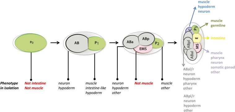
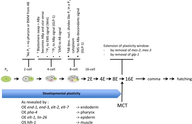
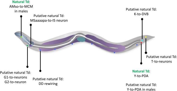
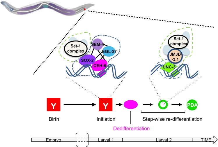
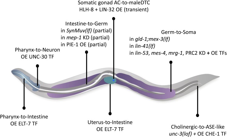
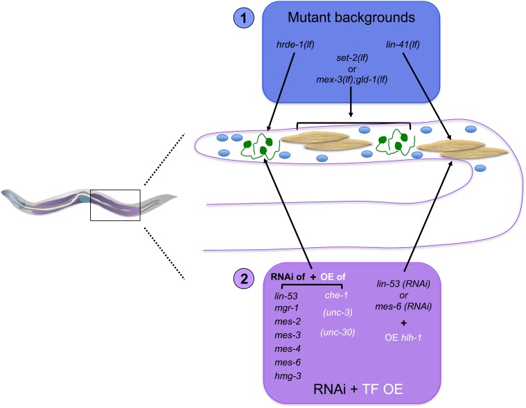
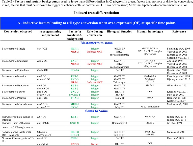
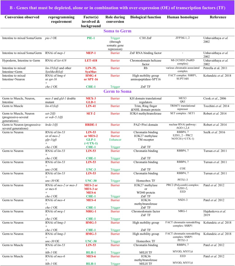
While Caenorhabditis elegans was originally regarded as a model for investigating determinate developmental programs, landmark studies have subsequently shown that the largely invariant pattern of development in the animal does not reflect irreversibility in rigidly fixed cell fates. Rather, cells at all stages of development, in both the soma and germline, have been shown to be capable of changing their fates through mutation or forced expression of fate-determining factors, as well as during the normal course of development. In this chapter, we review the basis for natural and induced cellular plasticity in C. elegans We describe the events that progressively restrict cellular differentiation during embryogenesis, starting with the multipotency-to-commitment transition (MCT) and subsequently through postembryonic development of the animal, and consider the range of molecular processes, including transcriptional and translational control systems, that contribute to cellular plasticity. These findings in the worm are discussed in the context of both classical and recent studies of cellular plasticity in vertebrate systems.

Keywords: WormBook; cell type conversion; reprogramming; stem cells; transdetermination; transdifferentiation.


Figures

Figure 1
Figure 1
Early embryonic lineage, evidence for cell-intrinsic determination and nonlinear segregation of developmental potential. The fertilized egg, named P0, divides unequally into the AB and P1 blastomeres. AB further divides into ABa and ABp, which will follow different division patterns. The tissue contribution of their descendants is indicated in gray. P1 divides unequally to give rise to the EMS and P2 blastomeres. P2 will give rise to the C and P3 blastomeres, and EMS gives rise to MS and E. The tissue contributions of the descendants of the C, P3, E, and MS blastomeres is indicated on the right. The cellular phenotype observed (in black) or not (in red) after culture in isolation, with or without cell arrest, of each blastomere up to the four-cell stage is also indicated.
Figure 2
Figure 2
Evidence for the early developmental plasticity window and the control of its timing. Early blastomeres up to the 8E-cell stage can exhibit high plasticity, as revealed by blastomeres swap (§), blastomeres ablations or culture in isolation (*), nuclear transfer (&) or analysis of lineage mutants (£). In addition, overexpression (OE) of defined TFs during this time-window transforms most, if not all, blastomeres into cells bearing characteristics of the indicated tissues. Transition from the 8E to the 16E-cell stage marks the multipotency-to-commitment transition (MCT). The window can be extended by removing the polycomb PRC2 complex (mes-2, mes-3 mutants) or early Notch activity (glp-1 mutant).
Figure 3
Figure 3
Schematic representation of the demonstrated and putative natural transdifferentiations in the worm. The initial and final identities, as well as their approximate localization in the worm are shown. Blue, pharynx; dark gray, intestine; purple, germline; green, uterus. Individual cells are represented as colored dots; red: rectal Y cell, green: amphid socket (AMso), in the male; dark green: G1 and G2 excretory pore cells; turquoise: rectal K cell; blue, DD neurons; dark blue: MSaaaapa cell; yellow: phasmid socket T cell. Anterior is to the left, and ventral to the bottom.
Figure 4
Figure 4
Rectal-to-neuron transdifferentiation. Rectal Y-to-PDA neuron Td occurs through a succession of discrete steps, including a dedifferentiation step, where the cell is stripped of its initial rectal identity without acquiring more cellular potential, followed by apparent step-wise redifferentiation into the PDA neuron. This succession of cellular steps is mirrored at the molecular level by step-specific combinations of histone modifier complexes (outlined in dashed green), such as JMJD-3.1 and the SET-1/MLL complex, and transcription factor complexes (outlined in dashed blue) such as a NODE-like + SOX-2 complex, and the UNC-3/COE TF.
Figure 5
Figure 5
Schematic representation of the known experimentally induced transdifferentiations in the worm. The initial and final identities, as well as their approximate localization in the worm are shown. Blue, pharynx; dark gray, intestine; purple, germline; green, uterus, blue dot, somatic gonad AC cell. lf, loss-of-function; KD, RNAi-mediated knock down; OE, overexpression; TF, transcription factor. Anterior is to the left, and ventral to the bottom.
Figure 6
Figure 6
Germ-to-soma transdifferentiation. Translational repressors and chromatin complexes safeguard the C. elegans germline identity: (1) In mutants for translational repressors or H3K4 methylation, listed in the blue box, germ cells (blue ovals) adopt several different somatic fates and resemble neuron (green cells) or muscle (beige cells)-like cells. Note that the germ-to-soma conversion seen in set-2 and hrde-1 mutants, members of the SET1 complex, arises progressively over several generations. (2) After knock down by RNAi of one of the following chromatin factors [lin-53, mes-2, -3, -4, -6, mrg-1, hmg-3; purple box], overexpression (OE) of a developmental regulator transcription factor (TF; white lettering) leads to specific germ-to-soma conversion: che-1 OE leads to ASE-like neuronal fate; unc-30 OE to a GABAergic neuronal-like fate; unc-3 OE to a cholinergic neuronal-like fate and hlh-1 OE leads to a muscle-like fate.
None
None

Similar articles

Cited by

References

    1. Abdus-Saboor I., Mancuso V. P., Murray J. I., Palozola K., Norris C. et al. , 2011. Notch and Ras promote sequential steps of excretory tube development in C. elegans. Development 138: 3545–3555. 10.1242/dev.068148 - DOI - PMC - PubMed
    1. Andersen E. C., Gerke J. P., Shapiro J. A., Crissman J. R., Ghosh R. et al. , 2012. Chromosome-scale selective sweeps shape Caenorhabditis elegans genomic diversity. Nat. Genet. 44: 285–290. 10.1038/ng.1050 - DOI - PMC - PubMed
    1. Andralojc K. M., Campbell A. C., Kelly A. L., Terrey M., Tanner P. C. et al. , 2017. ELLI-1, a novel germline protein, modulates RNAi activity and P-granule accumulation in Caenorhabditis elegans. PLoS Genet. 13: e1006611 10.1371/journal.pgen.1006611 - DOI - PMC - PubMed
    1. Ang Y. S., Tsai S. Y., Lee D. F., Monk J., Su J. et al. , 2011. Wdr5 mediates self-renewal and reprogramming via the embryonic stem cell core transcriptional network. Cell 145: 183–197. 10.1016/j.cell.2011.03.003 - DOI - PMC - PubMed
    1. Ashe A., Sapetschnig A., Weick E. M., Mitchell J., Bagijn M. P. et al. , 2012. piRNAs can trigger a multigenerational epigenetic memory in the germline of C. elegans. Cell 150: 88–99. 10.1016/j.cell.2012.06.018 - DOI - PMC - PubMed

Publication types

MeSH terms