. 2019 Nov 19;116(47):23772-23782.
doi: 10.1073/pnas.1913092116. Epub 2019 Nov 4.

The generation and propagation of the human alpha rhythm

Affiliations

The generation and propagation of the human alpha rhythm

Mila Halgren et al. Proc Natl Acad Sci U S A. .

Abstract

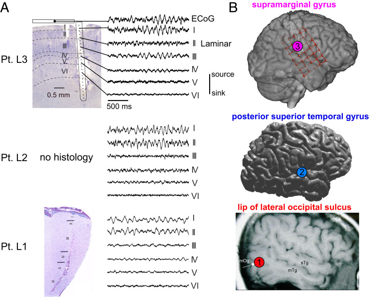
The alpha rhythm is the longest-studied brain oscillation and has been theorized to play a key role in cognition. Still, its physiology is poorly understood. In this study, we used microelectrodes and macroelectrodes in surgical epilepsy patients to measure the intracortical and thalamic generators of the alpha rhythm during quiet wakefulness. We first found that alpha in both visual and somatosensory cortex propagates from higher-order to lower-order areas. In posterior cortex, alpha propagates from higher-order anterosuperior areas toward the occipital pole, whereas alpha in somatosensory cortex propagates from associative regions toward primary cortex. Several analyses suggest that this cortical alpha leads pulvinar alpha, complicating prevailing theories of a thalamic pacemaker. Finally, alpha is dominated by currents and firing in supragranular cortical layers. Together, these results suggest that the alpha rhythm likely reflects short-range supragranular feedback, which propagates from higher- to lower-order cortex and cortex to thalamus. These physiological insights suggest how alpha could mediate feedback throughout the thalamocortical system.

Keywords: alpha; intracranial EEG; laminar; oscillations; thalamocortical.


Conflict of interest statement

The authors declare no competing interest.

Figures

Fig. 1.
Fig. 1.
Analysis stream for ECoG traveling waves. (A) Alpha propagates as a traveling wave in the raw broadband data. (B) Analysis stream for visualizing traveling waves in ECoG. We start with raw alpha phase of the grid over time. Then, for each time point, we find the circular distance (distance on the unit circle) between each contact and the grid’s mean phase (across all contacts) at that time point. Lastly, we find the circular mean of this difference to get each contact’s average phase advance or delay.
Fig. 2.
Fig. 2.
Alpha propagates from anterosuperior to posteroinferior cortex. (A) Alpha-phase snapshots from Pt. L1 demonstrate propagation from the grid’s top-right (anterosuperior) to bottom-left corner (posteroinferior). (B) Average circular distance of each contact’s alpha phase from the spatial mean phase during eye closure. In all patients, alpha propagates toward posteroinferior areas. Overlaid arrow is the direction of the grid’s average phase gradient. Color runs from ±π3 in Pts. E1, E2, and E5 and macaque; and from ±π5 in Pts. E3 and E4. (C) Average probability distribution of traveling wave directions across time such that the bottom left contact is the most posteroinferior (±SEM across patients). (D) Average probability distribution of traveling wave speeds (±SEM across patients).
Fig. 3.
Fig. 3.
Robust alpha rhythms can be recorded in human pulvinar and cortex. (A) Representative 6-s LFPg traces of simultaneous thalamic and cortical alpha activity. Prominent, largely continuous alpha rhythms can be recorded in various locations within the pulvinar as well as posterior cortex. (B) Cortical implant locations in all SEEG patients displayed on Pt. S3′s brain. Each color signifies a different patient.
Fig. 4.
Fig. 4.
Cortical alpha leads thalamic (pulvinar) alpha. (A) Average alpha-phase lag/leads in bipolar contacts (n = 5). Note that anterosuperior channels lead inferoposterior ones, in accord with our ECoG recordings. (B) Power spectra of the thalamic (color) and cortical (gray) channel with the greatest alpha power. (C) The difference in start times between all cortical and thalamic alpha bursts (start time in thalamus to start time in cortex) in the 28 channel pairs with a significant thalamic or cortical lead (P < 0.05, Bonferroni corrected, binomial test). Alpha bursts start (on average) in cortex for all 28 channel pairs. (D) Cortical and thalamic LFPg and HGP from representative channels locked to peaks in thalamic alpha LFPg—cortical, but not thalamic, HGP is phasic with thalamic LFPg. (E) Coherence spectra of thalamic and cortical LFPg with thalamic and cortical HGP and LFPg from the same channel pair in D; the coherence of thalamic LFPg with cortical HGP (but not thalamic HGP) suggests that the cortex may drive thalamic alpha activity. (F) Normalized thalamic and cortical HGP at different thalamic alpha phases averaged across channels; note that cortical HGP slightly leads thalamic HGP. (G) GC spectra averaged over all thalamocortical contact pairs; corticothalamic causality shows a strong alpha peak. Amp., amplitude; a.u., arbitrary units; ctx, cortical; norm., normalized; thal, thalamic.
Fig. 5.
Fig. 5.
Laminar recordings of cortical alpha. (A) Nissl stains of the explanted tissue surrounding the laminar probe in Pts. L1 and L3, in addition to representative laminar CSD traces from each layer in each subject. Note that despite being made in distinct cortical locations, alpha oscillations were always strongest in layer I/II. Furthermore, in L3, the trace of a simultaneously recorded overlying ECoG contact is near identical to the underlying laminar layer I. (B) Locations of each laminar probe in all patients. Adapted with permission from ref. .
Fig. 6.
Fig. 6.
Alpha CSD and HGP are maximal in supragranular cortex. (A) Average CSD and HGP waveforms of a single channel on the same time axes as B (±SEM across alpha sinks). (B and C) CSD (B) and HGP (C) averaged on current sinks in channels 3 and 6 in Pts. L2 and L3, respectively; white and black dashed lines indicate layer IV boundaries and the time of the alpha sink, respectively. (D) Z score of the MI between alpha phase and HGP across all channels (Ch.). (E) Average alpha power throughout the cortical depth (±SEM across epochs). (F) Power spectra of the channel with greatest alpha power in each subject (±SEM across epochs).
Fig. 7.
Fig. 7.
MUA is modulated in layer III. (A) MUA averaged on current sinks on the channels with the greatest alpha power (channels 3 and 6, respectively), which is most clearly modulated within lower layer III. (B) Tort’s MI between alpha CSD phase and MUA amplitude over all laminar contact pairs—note that firing is correlated with alpha phase in both superficial and deep cortex. Ch., channel.
Fig. 8.
Fig. 8.
Simultaneous ECoG–laminar recordings reveal traveling alpha waves which propagate through supragranular cortex. (A) Average circular distance of each ECoG (circles) and layer I laminar (diamond) contact’s alpha phase from the spatial mean phase throughout the ECoG grid. Note that the laminar’s alpha phase is intermediate to neighboring ECoG contacts, suggesting that ECoG and the laminar probe recording the same traveling wave at different scales. (B) Representative drawing of a traveling alpha wave (as measured with ECoG) propagating through superficial layers (as measured by a laminar probe). (C) Example traces from ECoG contacts posterior (red) and anterior (blue) to the laminar probe. Alpha phase in the laminar is intermediary to the ECoG contacts. (D) Distribution of traveling wave directions; mu waves propagate from posterior (higher-order) toward anterior (lower-order) cortex. (E) Power spectra from simultaneous laminar and ECoG recordings; they share a near-identical alpha peak. (F) Laminar CSD averaged on troughs in the nearest ECoG contact. Note that alpha activity is superficial.
Fig. 9.
Fig. 9.
Tentative model for how alpha’s physiology could mediate feedback. (A) Alpha propagates as a traveling wave from higher-order (middle temporal, visual area 3) toward lower-order visual areas 1/2 cortical areas. (B) Alpha is strongest within supragranular cortex and may carry top-down information via short-range feedback connections to constrain lower-level processing; for instance, alpha may play a role in resolving ambiguous visual imagery, such as the picture of a woman and a horse’s snout shown above. Cortical alpha in layer VI might influence alpha activity within the pulvinar. Reproduced with permission from ref. , which is licensed under CC BY 4.0.

References

    1. Berger H., Das elektrenkephalogramm des menschen. Naturwissenschaften 23, 121–124 (1935).
    1. Ito J., Nikolaev A. R., van Leeuwen C., Spatial and temporal structure of phase synchronization of spontaneous alpha EEG activity. Biol. Cybern. 92, 54–60 (2005). - PubMed
    1. von Stein A., Sarnthein J., Different frequencies for different scales of cortical integration: From local gamma to long range alpha/theta synchronization. Int. J. Psychophysiol. 38, 301–313 (2000). - PubMed
    1. Saalmann Y. B., Pinsk M. A., Wang L., Li X., Kastner S., The pulvinar regulates information transmission between cortical areas based on attention demands. Science 337, 753–756 (2012). - PMC - PubMed
    1. Samaha J., Postle B. R., The speed of alpha-band oscillations predicts the temporal resolution of visual perception. Curr. Biol. 25, 2985–2990 (2015). - PMC - PubMed

Publication types

LinkOut - more resources