

Functional Connectome of the Fetal Brain
- PMID: 31685648
- PMCID: PMC6891066
- DOI: 10.1523/JNEUROSCI.2891-18.2019
Functional Connectome of the Fetal Brain
Abstract
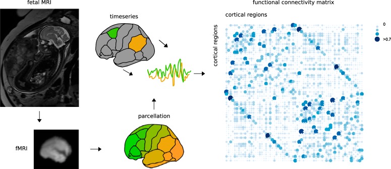
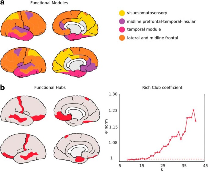
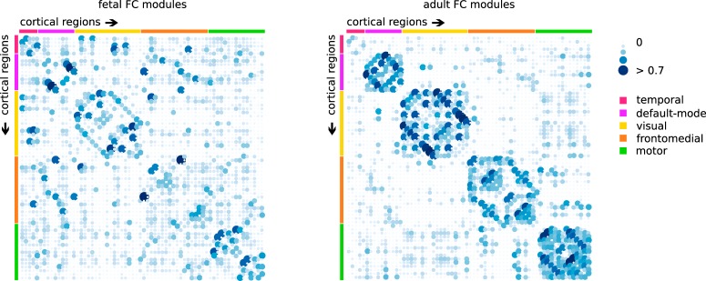
Large-scale functional connectome formation and reorganization is apparent in the second trimester of pregnancy, making it a crucial and vulnerable time window in connectome development. Here we identified which architectural principles of functional connectome organization are initiated before birth, and contrast those with topological characteristics observed in the mature adult brain. A sample of 105 pregnant women participated in human fetal resting-state fMRI studies (fetal gestational age between 20 and 40 weeks). Connectome analysis was used to analyze weighted network characteristics of fetal macroscale brain wiring. We identified efficient network attributes, common functional modules, and high overlap between the fetal and adult brain network. Our results indicate that key features of the functional connectome are present in the second and third trimesters of pregnancy. Understanding the organizational principles of fetal connectome organization may bring opportunities to develop markers for early detection of alterations of brain function.SIGNIFICANCE STATEMENT The fetal to neonatal period is well known as a critical stage in brain development. Rapid neurodevelopmental processes establish key functional neural circuits of the human brain. Prenatal risk factors may interfere with early trajectories of connectome formation and thereby shape future health outcomes. Recent advances in MRI have made it possible to examine fetal brain functional connectivity. In this study, we evaluate the network topography of normative functional network development during connectome genesis in utero Understanding the developmental trajectory of brain connectivity provides a basis for understanding how the prenatal period shapes future brain function and disease dysfunction.
Keywords: brain development; fetal; functional connectivity; prenatal; resting-state fMRI.
Copyright © 2019 the authors.
Figures
Comment in
-
The Fetal Functional Connectome Offers Clues for Early Maturing Networks and Implications for Neurodevelopmental Disorders.J Neurosci. 2020 Jun 3;40(23):4436-4438. doi: 10.1523/JNEUROSCI.0260-20.2020. J Neurosci. 2020. PMID: 32493796 Free PMC article. No abstract available.
-
Neural Mechanisms for Prediction: From Action to Higher-Order Cognition.J Neurosci. 2020 Jul 1;40(27):5158-5160. doi: 10.1523/JNEUROSCI.0732-20.2020. J Neurosci. 2020. PMID: 32611592 Free PMC article. No abstract available.
-
Adaptive Immune Cells Link Microbial Metabolites to Stroke Recovery.J Neurosci. 2020 Jul 8;40(28):5344-5346. doi: 10.1523/JNEUROSCI.0634-20.2020. J Neurosci. 2020. PMID: 32641452 Free PMC article. No abstract available.
References
-
- Ashburner J, Barnes G, Chen C, Daunizeau J, Flandin G, Friston K, Gitelman D, Kiebel S, Kilner J, Litvak V (2012) SPM8 manual. London UK: Functional Imaging Laboratory, Institute of Neurology.
Publication types
MeSH terms
Grants and funding
LinkOut - more resources
Full Text Sources