. 2019 Nov 4;10(11):830.
doi: 10.1038/s41419-019-2054-7.

ASPP2 inhibits tumor growth by repressing the mevalonate pathway in hepatocellular carcinoma

Affiliations

ASPP2 inhibits tumor growth by repressing the mevalonate pathway in hepatocellular carcinoma

Beibei Liang et al. Cell Death Dis. .

Abstract

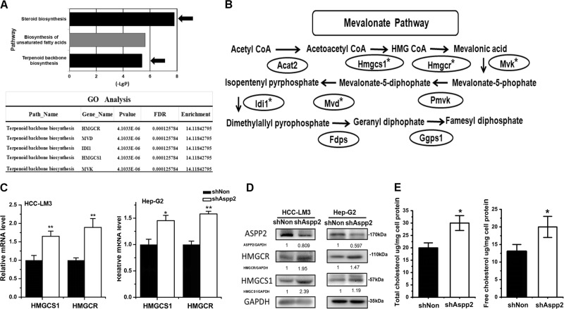
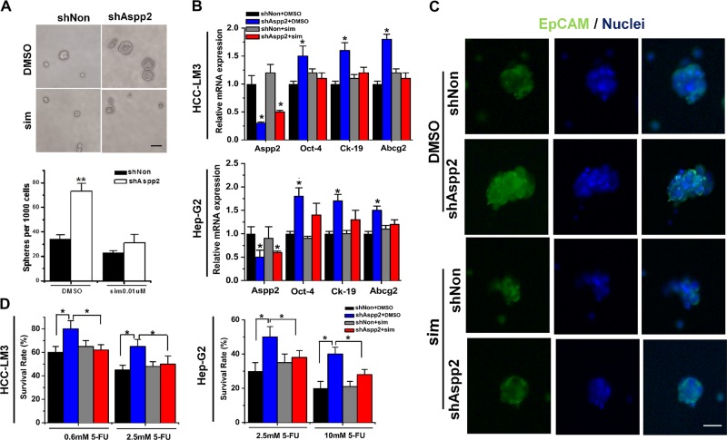
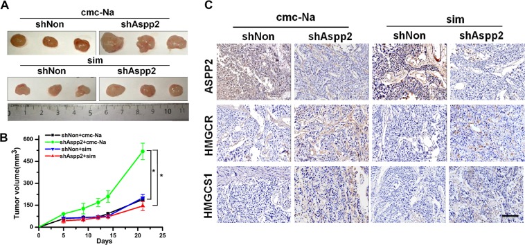
Cancer is, fundamentally, a disorder of cell growth and proliferation, which requires adequate supplies of energy and nutrients. In this study, we report that the haplo-insufficient tumor suppressor ASPP2, a p53 activator, negatively regulates the mevalonate pathway to mediate its inhibitory effect on tumor growth in hepatocellular carcinoma (HCC). Gene expression profile analysis revealed that the expression of key enzymes in the mevalonate pathway were increased when ASPP2 was downregulated. HCC cells gained higher cholesterol levels and enhanced tumor-initiating capability in response to the depletion of ASPP2. Simvastatin, a mevalonate pathway inhibitor, efficiently abrogated ASPP2 depletion-induced anchorage-independent cell proliferation, resistance to chemotherapy drugs in vitro, and tumor growth in xenografted nude mice. Mechanistically, ASPP2 interacts with SREBP-2 in the nucleus and restricts the transcriptional activity of SREBP-2 on its target genes, which include key enzymes involved in the mevalonate pathway. Moreover, clinical data revealed better prognosis in patients with high levels of ASPP2 and low levels of the mevalonate pathway enzyme HMGCR. Our findings provide functional and mechanistic insights into the critical role of ASPP2 in the regulation of the mevalonate pathway and the importance of this pathway in tumor initiation and tumor growth, which may provide a new therapeutic opportunity for HCC.


Conflict of interest statement

The authors declare that they have no conflict of interest.

Figures

Fig. 1
Fig. 1. Downregulation of ASPP2 enhances mevalonate pathway gene expression and cholesterol biosynthesis in HCC cells.
a Top: KEGG Pathway analysis of ASPP2 knockdown show significant differential changes (P < 0.05) in canonical metabolic pathways. The P value for each pathway is indicated by the bar and is expressed as −1 times the log of the P value. Arrows are used to indicated the two pathways closely related with mevalonate metabolism. Bottom: Gene Ontology (GO) analysis revealed genes were most significantly upregulated and enriched by ASPP2 knockdown in the terpenoid backbone biosynthesis pathway (P < 0.05, FDR < 0.25). b Schematic of the mevalonate pathway with asterisk (*) marking upregulated genes by ASPP2 knockdown in panel (a) bottom. c qRT-PCR of Hmgcs1, Hmgcr mRNA expression in HCC-LM3 and HepG2 cells infected with lentivirus encoding shRNA was indicated for 72 h. β-actin was used as a control. d Corresponding western-blot of HMGCS1 and HMGCR in HCC-LM3 and HepG2 cells. e Total cholesterol and free cholesterol were measured by GC-MS methods, using the internal standard strategy as methods described. Each symbol represents the mean ± SD of three wells. Asterisk (*) indicates P < 0.05
Fig. 2
Fig. 2. Mevalonate metabolism is essential for maintaining tumor-initiating capability in ASPP2-depleted HCC cells.
a Top: Representative images of spheroid formation in HCC-LM3 cells treated with 0.01 μM simvastatin or DMSO after knock down of ASPP2. Bottom: the number of spheres formation per 1000 HCC-LM3 cells.Scale bar, 100 mm. b qRT-PCR gene expression of Aspp2, Oct-4, Ck-19, and Abcg2 in HCC-LM3 and HepG2 cells transfected with the indicated lentivirus and treated with 10 μM simvastatin or DMSO. c Immunofluorescence stainings of HCC-LM3 spheres formed from cells transfected with the indicated lentivirus and treated with 10 μM simvastatin or DMSO. Green staining represented EpCAM protein. DAPI was used stain nuclear(blue staining) Scale bar, 40 mm d Cell viability of the infected HCC-LM3 and HepG2 cell treated with 10 μM simvastatin or DMSO and 5-FU(0.6 mM,2.5 mM or 10 mM) was analyzed by MTS assay. Asterisk (*) indicates P < 0.05, asterisks (**) indicates P < 0.01
Fig. 3
Fig. 3. Downregulation of ASPP2 promoted tumor growth in vivo by activating mevalonate biosynthesis.
a Representative dissected tumors from nude mice treated without (top) or with (bottom) simvastatin for 3 weeks and corresponding volume measurement, b Asterisk (*) indicates P < 0.05. c Immunohistochemical staining of tumors derived from nude mice for ASPP2, HMGCR, and HMGCS1 (magnification, ×200). ASPP2-silenced groups exhibited increased HMGCR and HMGCS-1 expression. These alterations were compromised in ASPP2-silenced groups treated with simvastatin
Fig. 4
Fig. 4. Downregulation of ASPP2 promotes mevalonate metabolism by activation of SREBP-2.
a Immunofluorescent images of HCC-LM3 cells showing the cellular location of endogenous ASPP2 and SREBP-2. ASPP2 and SREPB-2 were stained green and red, respectively. DAPI (blue stain) was used to stain the nucleas.Scale bars: 40 μm. b Western blot of the immunoprecipitated ASPP2-SREBP-2 complex in total cell lysates (Left) or nuclear proteins lysates (Right) with anti-ASPP2 in HCC-LM3 cells. c Western blots of V5-tagged ASPP2 co-transfected with HA-SREBP-2 into HEK293T following IP with anti-V5 antibody in total cell lysates (Left) and nuclear proteins lysates (Right). d Western blots of HA-tagged SREBP-2 co-transfected with ASPP2-V5 into HEK293T following IP with anti-HA antibody in total cell lysates (Left) and nuclear proteins lysates (Right). e Luciferase activity of Hamster HMGCS1, human LDLR promoters and three Sterol regulatory elements (3SRE) with respect to reporter vector pGL3.0. The constructs were co-transfected with an internal control pRL-TK vector into HCC-LM3 cells with silencing of ASPP2 (left) and Huh-7 cell with ASPP2 overexpression (right). f The number of spheres per 1000 HCC cells with siRNA negative control or siRNA Srebp-2 after knock down of ASPP2. g Western blot of HMGCR and HMGCS1 protein in HCC-LM3 cells infected with LV-shASPP2 or LV-shNon and treated with siAspp2 and/or siSrebp2. Asterisk (*) indicates P < 0.05, asterisks (**) indicates P < 0.01
Fig. 5
Fig. 5. Expression of ASPP2 correlates negatively with HMGCR in surgical specimens of HCC.
a ASPP2 and HMGCR were detected by immunohistochemical staining in 80 HCC samples. Representative pictures are shown for four patients. (Scale bars: 50 μm). b Table of ASPP2 and HMGCR high and low expression according to immunohistochemistry scoring. Significant negative correlation between ASPP2 and HMGCR expression was found, Fisher’s test P < 0.05. c The strong expression of ASPP2 in nuclear in 17 HCC samples. Representative pictures of immunohistochemical staining are shown for two patients.Scale bars: 50 μm. d. Table of positive ASPP2 expression in nuclear or cytoplasm and their HMGCR expression according to the scores of immunohistochemistry staining. e Left: Overall survival (OS) and right: recurrence-free survival (RFS) rate curves of the analyzed four subgroups (ASPP2 low/HMGCR low; ASPP2 low/HMGCR high; ASPP2 high /HMGCR low; ASPP2 high/HMGCR high), (n = 80). Kaplan-Meier analysis showed RFS (P < 0.05) and OS (P < 0.05) were significantly best among patients with ASPP2-high and HMGCR-low expression

References

    1. Hanahan D, Weinberg RA. Hallmarks of cancer: the next generation. Cell. 2011;144:646–674. doi: 10.1016/j.cell.2011.02.013. - DOI - PubMed
    1. Simons K, Ikonen E. How cells handle cholesterol. Science. 2000;290:1721–1726. doi: 10.1126/science.290.5497.1721. - DOI - PubMed
    1. Beloribi-Djefaflia S, Vasseur S, Guillaumond F. Lipid metabolic reprogramming in cancer cells. Oncogenesis. 2016;5:e189. doi: 10.1038/oncsis.2015.49. - DOI - PMC - PubMed
    1. Kuzu OF, Noory MA, Robertson GP. The role of cholesterol in cancer. Cancer Res. 2016;76:2063–2070. doi: 10.1158/0008-5472.CAN-15-2613. - DOI - PMC - PubMed
    1. Murtola TJ, et al. The importance of LDL and cholesterol metabolism for prostate epithelial cell growth. PloS ONE. 2012;7:e39445. doi: 10.1371/journal.pone.0039445. - DOI - PMC - PubMed

Publication types

MeSH terms