. 2019 Nov 4;10(11):834.
doi: 10.1038/s41419-019-2066-3.

The paracrine induction of prostate cancer progression by caveolin-1

Affiliations

The paracrine induction of prostate cancer progression by caveolin-1

Chun-Jung Lin et al. Cell Death Dis. .

Abstract

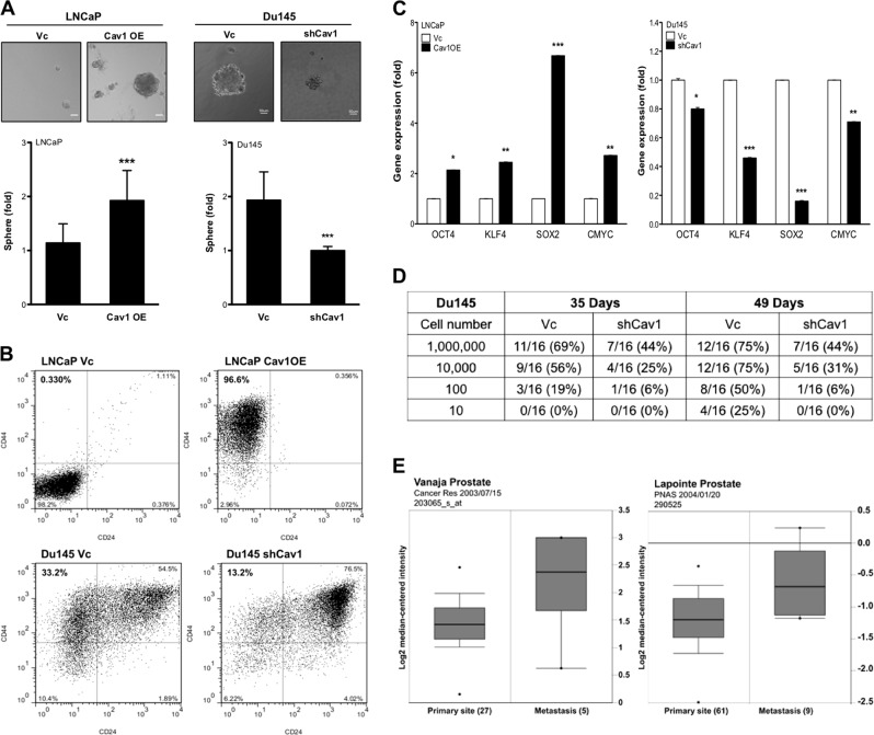
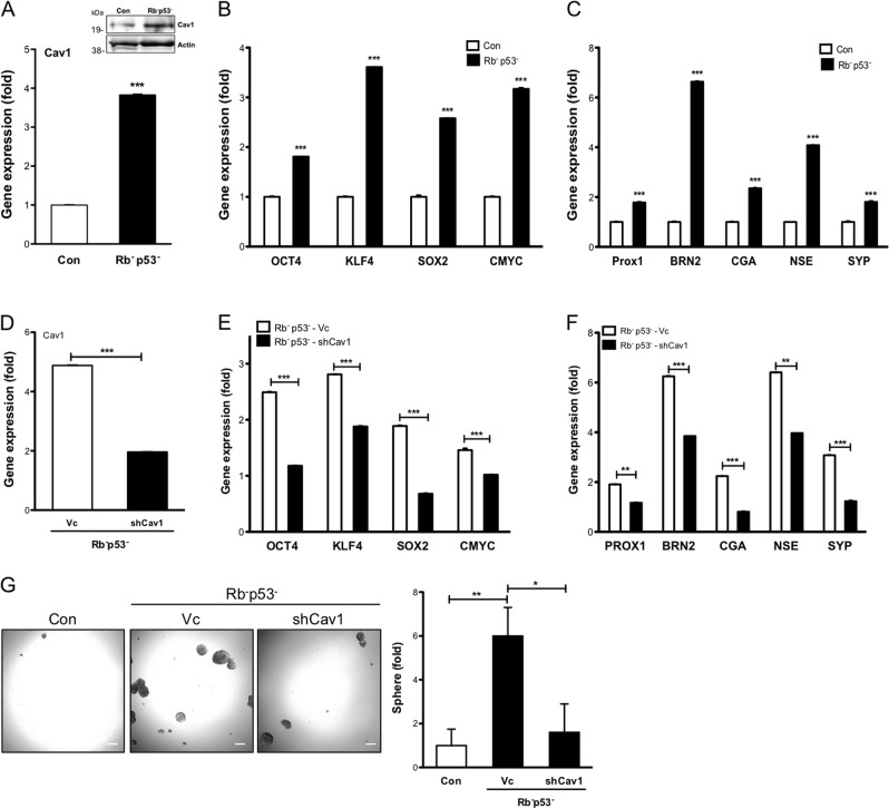
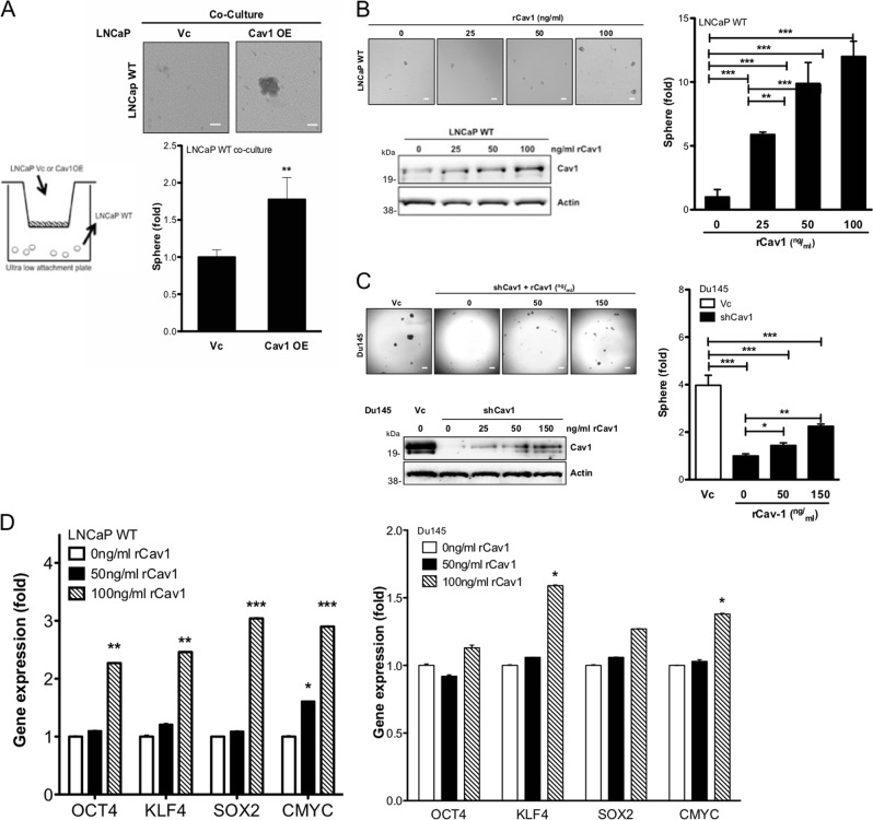
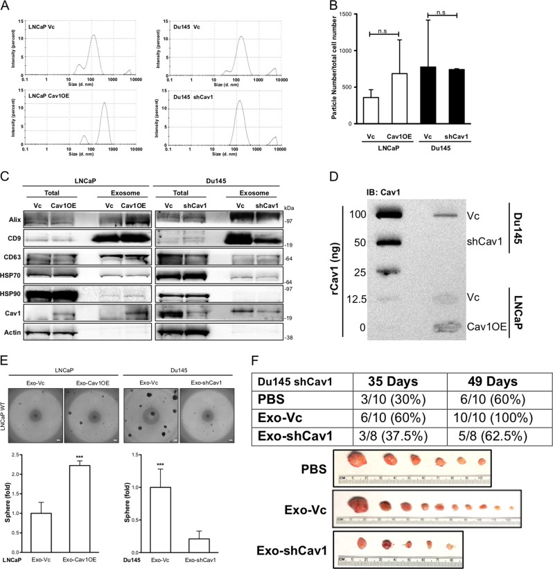
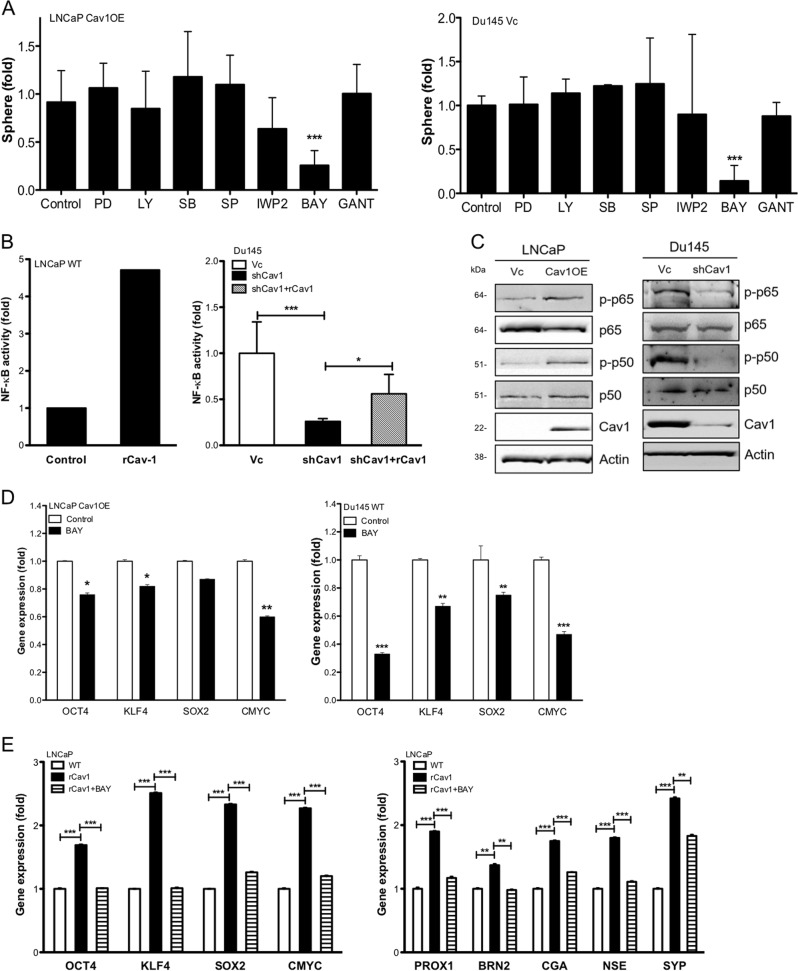
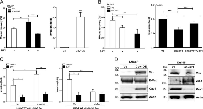
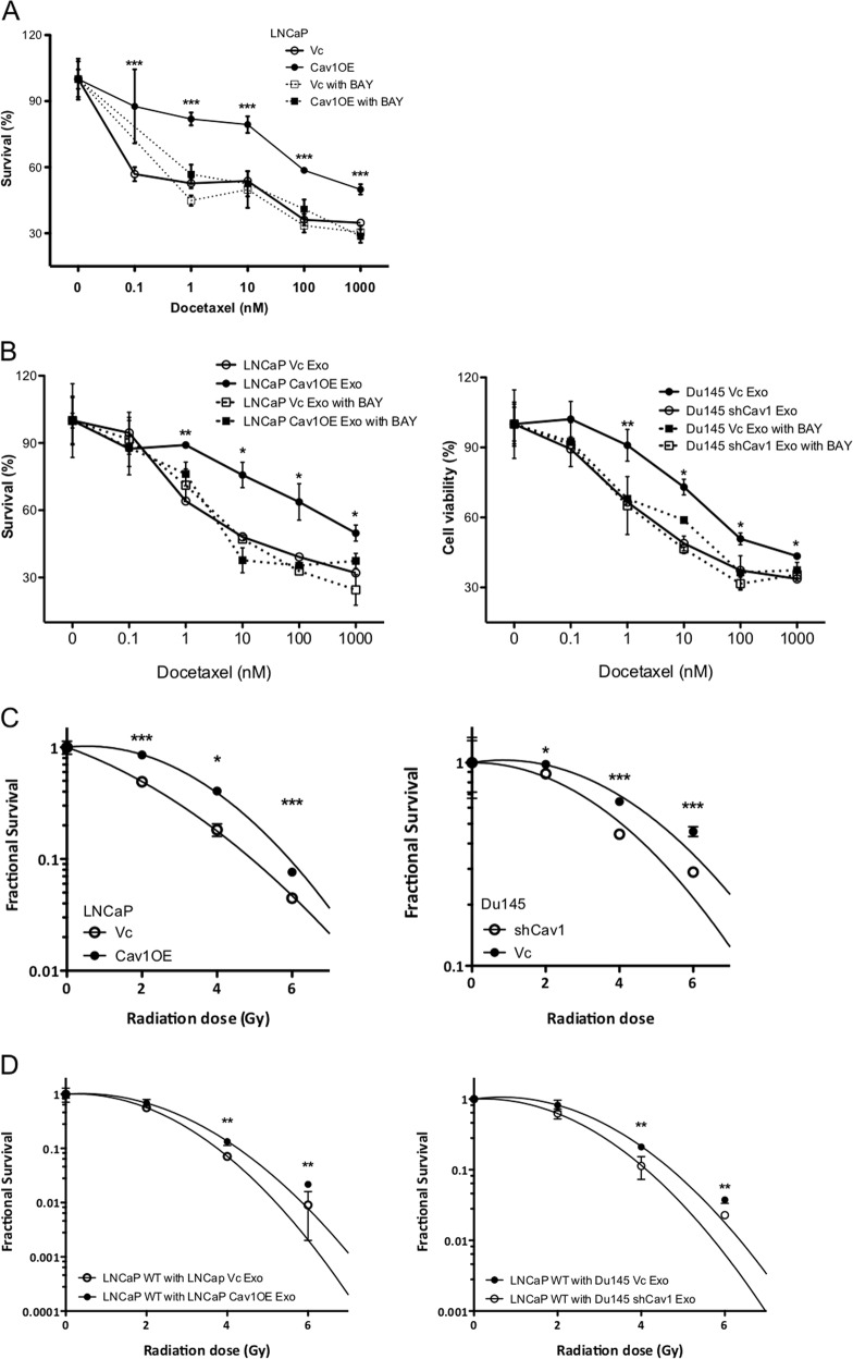
A subpopulation of cancer stem cells (CSCs) plays a critical role of cancer progression, recurrence, and therapeutic resistance. Many studies have indicated that castration-resistant prostate cancer (CRPC) is associated with stem cell phenotypes, which could further promote neuroendocrine transdifferentiation. Although only a small subset of genetically pre-programmed cells in each organ has stem cell capability, CSCs appear to be inducible among a heterogeneous cancer cell population. However, the inductive mechanism(s) leading to the emergence of these CSCs are not fully understood in CRPC. Tumor cells actively produce, release, and utilize exosomes to promote cancer development and metastasis, cancer immune evasion as well as chemotherapeutic resistance; the impact of tumor-derived exosomes (TDE) and its cargo on prostate cancer (PCa) development is still unclear. In this study, we demonstrate that the presence of Cav-1 in TDE acts as a potent driver to induce CSC phenotypes and epithelial-mesenchymal transition in PCa undergoing neuroendocrine differentiation through NFκB signaling pathway. Furthermore, Cav-1 in mCRPC-derived exosomes is capable of inducing radio- and chemo-resistance in recipient cells. Collectively, these data support Cav-1 as a critical driver for mCRPC progression.


Conflict of interest statement

The authors declare that they have no conflict of interest.

Figures

Fig. 1
Fig. 1. Cav-1 increases CSC subpopulation.
a Prostate sphere formation of LNCaP (Vc vs. CavOE) and Du145 (Vc vs. shCav1) was determined after 14 days cultured in ultralow attachment plate. Number of spheres was counted and normalized with control cells (e.g., LNCaP Vc or Du145 shCav1), respectively. Data were shown as mean ± SD (n > 10), ***P < 0.001. Scale bar = 50 μm. b CSC subpopulation was determined by Flow cytometry based on CD24low and CD44high population. c The gene expression of Yamanaka factors was measured by qRT-PCR. Data were shown as mean ± SD (n > 10), *P < 0.5, **P < 0.01, ***P < 0.001. e In vivo tumorigenicity of Du145 Vc vs. shCav1 cells was measured at Day 35 and Day 49 post injection. d A serial dilution of Du145 Vc or shCav1 cells was subcutaneously injected into immune-deficient mice. Tumor incidence was analyzed after 35 days and 49 days of injection. e Clinical correlation of Cav-1 mRNA expression in PCa patients from two cohorts was obtained by using Oncomine Database. Cav-1 mRNA expression was compared between primary site and metastasis site of tumor
Fig. 2
Fig. 2. Induction of Cav-1 in PCa carrying p53 and Rb mutation correlates with CSC and NED-related gene expression.
The expression profile of a Cav-1 in mRNA and protein expression level, b Yamanaka factors (OCT4, KLF4, SOX2, CMYC), and c NED-related genes (PROX1, BRN2, CGA, NSE, SYP) were determined in LNCaP Con vs. LNCAP (p53, Rb) by qRT-PCR. The gene expression profile of d Cav-1, e Yamanaka factors genes (OCT4, KLF4, SOX2, CMYC), and f NED-related genes (PROX1, BRN2, CGA, NSE, SYP) were determined by qRT-PCR in LNCaP (p53, Rb) cells after Cav-1 knockdown using specific shRNA. g The sphere formation in LNCaP Con, LNCAP (p53, Rb)-Vc and LNCAP (p53, Rb)-shCav1 cells. Scale bar = 100 μm. All data were shown as mean ± SD (n > 10), *P < 0.5, **P < 0.01, ***P < 0.001
Fig. 3
Fig. 3. Exogenous rCav-1 promotes CSC phenotypes.
a Prostate sphere formation of WT LNCaP under the influence of Cav-1-expressing cells was determined after plating LNCaP WT (500 cells) onto the bottom of an ultralow attachment 24-well plate and either LNCaP Vc or Cav1OE (5 × 104 cells) onto a Transwell (3.0-µm polycarbonate). Number of spheres was determined 14 days after co-culture. Scale bar = 50 μm. b Upper panel: prostate sphere formation of LNCaP WT incubated with rCav-1 (from 0 to 100 ng/ml) was determined after 14 days. Lower panel: rCav-1 uptake by WT LNCaP was analyzed by western blot. Scale bar = 100 μm. c Upper panel: prostate sphere formation of Du145 shCav1 (low Cav-1 expression) incubated with rCav-1 (from 0 to 150 ng/ml) was determined after 14 days. Lower panel: rCav-1 uptake by Du145 shCav1was analyzed by western blot. Scale bar = 100 μm. d The gene expression profile of Yamanaka factors gene (Left panel) and NED-related genes (Right panel) was examined 48h post treatment. Scale bar = 50 μm. All data were shown as mean ± SD (n > 10), *P < 0.5, **P < 0.01, ***P < 0.001
Fig. 4
Fig. 4. Cav-1 delivered by exosomes promotes CSC phenotypes.
Characterization of exosomes purified from LNCaP (Vc or Cav1OE) or Du145 (Vc, shCav1) based on a particle size, b particle number per cell, and c exosome markers (Alix, CD9, CD63, HSP70, HSP90). d The amount of Cav-1 in purified exosomes was determined by dot blot assay. Cav-1 concentration was analyzed by rCav-1 (0–100 ng) standard curve. e Prostate sphere formation of WT LNCaP was determined after treating with purified exosomes from LNCaP (Vc or Cav1OE) or Du145 (Vc, shCav1) for 14 days of treatment. Scale bar = 100 μm. Data were shown as mean ± SD (n > 10), n.s., non-significant, *P < 0.5, **P < 0.01, ***P < 0.001. f Du145 shCav1 cells were subcutaneously injected in immune-deficient mice then treated with PBS or TDEs purified from Du145 Vc (Exo-Vc) or Du145 shCav1 (Exo-shCav1), twice-a-week and tumor incidence was recorded at Week 5 and Week 7 post injection. And all tumors were collected at Week 7
Fig. 5
Fig. 5. Cav-1 promotes CSC phenotypes through NFκB signaling pathway.
a The effect of small molecule inhibitors (PI3K/Akt: LY294002 (1 µm); Erk: PD98059 (2 µm), p38: SB202190 (100 nm); JNK: SP600125 (100 nm); Wnt: IWP2 (30 nm); NFκB: BAY11-7082 (5 µm); Gli1/2: GANT (5 µm) on prostate sphere formation of LNCaP Cav1OE and Du145 Vc cells. b The effect of rCav-1 on NFκB transcriptional activities was determined after transfecting luciferase reporter gene construct into WT LNCaP or Du145 (Vc or shCav1) cells. Relative luciferase activities were measured and then normalized with Renilla luciferase activities in each sample. c The activation status of NFκB pathway was determined by western blot. d The effect of NFκB inhibitor (BAY, 5 µm) on gene expression of Yamanaka factors in LNCaP Cav1OE or Du145 Vc cell was determined by qRT-PCR 24h after treatment. e The effect of NFκB inhibitor (BAY, 5 µm) on the expression of Yamanaka factors or NED-related genes in WT LNCaP cells treated with rCav-1 was determined by qRT-PCR. Data were shown as mean ± SD (n > 10), *P < 0.5, **P < 0.01, ***P < 0.001
Fig. 6
Fig. 6. Cav-1-elicited NFκB signaling pathway promotes cell migration, invasion, and EMT.
a The cell migratory or invasive abilities in LNCaP (Vc vs. Cav1OE) was measured using wound healing or Transwell invasion assay, respectively. b The cell migratory or invasive abilities in Du145 (Vc vs. shCav1) were measured using wound healing or Transwell invasion assay, respectively. c The effect of NFκB inhibitor (BAY, 5 µm) on cell invasion activities was measured in exosomes-treated WT LNCaP cell after 24h incubation. d The expression of EMT markers in LNCaP (Vc vs. Cav1OE) (Left panel) and Du145 (Vc vs. shCav1) (Right panel). All data were shown as mean ± SD (n > 10), *P < 0.5, **P < 0.01, ***P < 0.001
Fig. 7
Fig. 7. Cav-1 induces the resistance of PCa cells to chemotherapy or radiotherapy.
a The effect of NFκB inhibitor (BAY, 5 µm) on the sensitivity of LNCaP (Vc vs. Cav1OE) cells exposed to different concentrations of Docetaxel was determined by MTT assay 48 h post treatment. b The effect of NFκB inhibitor (BAY, 5 µm) on the sensitivity of WT LNCaP cells incubated with exosomes from various sources and different concentrations of Docetaxel was determined by MTT assay 48 h post treatment. c The effect of radiation (0–6 Gy) on cell survival of LNCaP (Vc vs. Cav1OE) or Du145 (Vc vs. shCav1) was determined 14 days post radiation. d The effect of radiation (0–6 Gy) on cell survival of WT LNCaP treated with exosomes from various sources was determined 14 days post radiation. All data were shown as mean ± SD (n > 10), *P < 0.5, **P < 0.01, ***P < 0.001

Similar articles

Cited by

References

    1. Siegel RL, Miller KD, Jemal A. Cancer statistics, 2016. CA Cancer J. Clin. 2016;66:7–30. doi: 10.3322/caac.21332. - DOI - PubMed
    1. Miller Kimberly D., Siegel Rebecca L., Lin Chun Chieh, Mariotto Angela B., Kramer Joan L., Rowland Julia H., Stein Kevin D., Alteri Rick, Jemal Ahmedin. Cancer treatment and survivorship statistics, 2016. CA: A Cancer Journal for Clinicians. 2016;66(4):271–289. - PubMed
    1. Smith RA, et al. Cancer screening in the United States, 2016: a review of current American Cancer Society guidelines and current issues in cancer screening. CA Cancer J. Clin. 2016;66:95–114. doi: 10.3322/caac.21336. - DOI - PubMed
    1. Tickner JA, Urquhart AJ, Stephenson SA, Richard DJ, O'Byrne KJ. Functions and therapeutic roles of exosomes in cancer. Front. Oncol. 2014;4:127. doi: 10.3389/fonc.2014.00127. - DOI - PMC - PubMed
    1. Mathivanan S, Ji H, Simpson RJ. Exosomes: extracellular organelles important in intercellular communication. J. Proteom. 2010;73:1907–1920. doi: 10.1016/j.jprot.2010.06.006. - DOI - PubMed

Publication types