. 2019 Nov 4;9(1):15954.
doi: 10.1038/s41598-019-52400-5.

CRISPR interference to interrogate genes that control biofilm formation in Pseudomonas fluorescens

Affiliations

CRISPR interference to interrogate genes that control biofilm formation in Pseudomonas fluorescens

Marie-Francoise Noirot-Gros et al. Sci Rep. .

Abstract

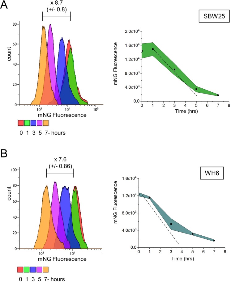
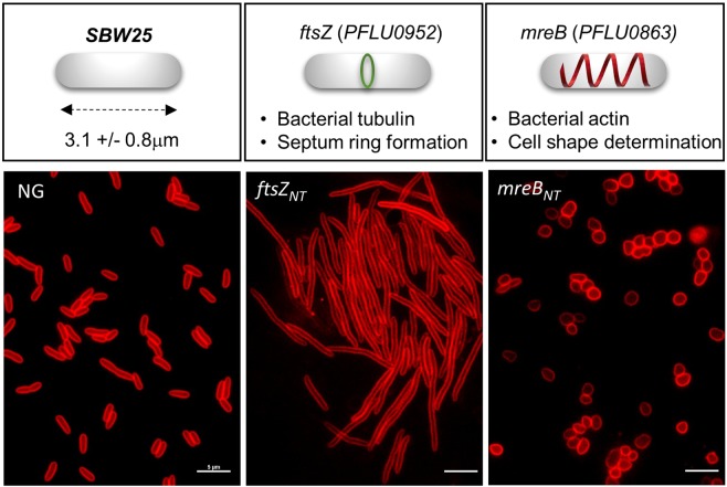
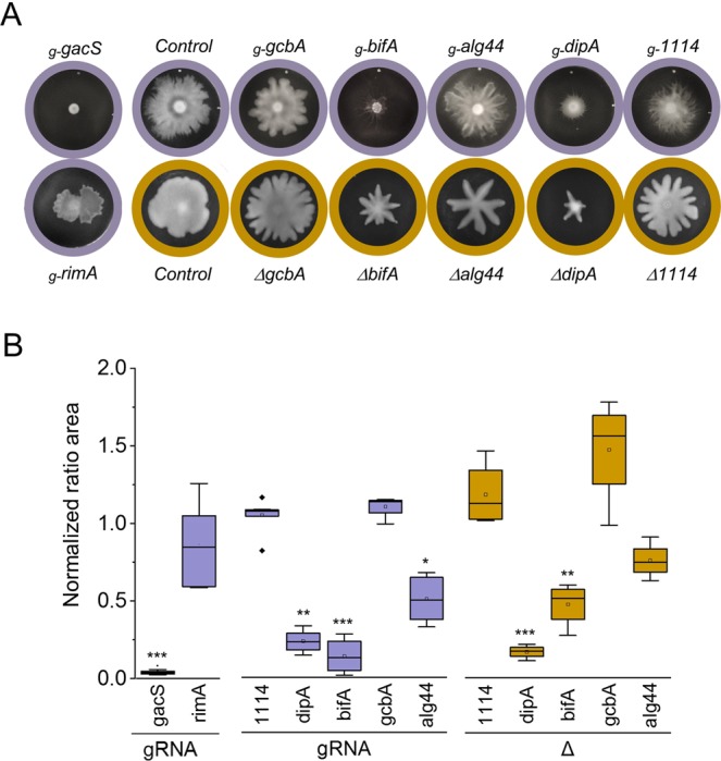
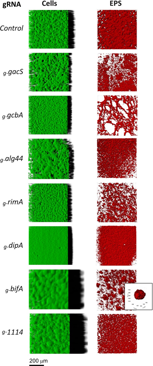
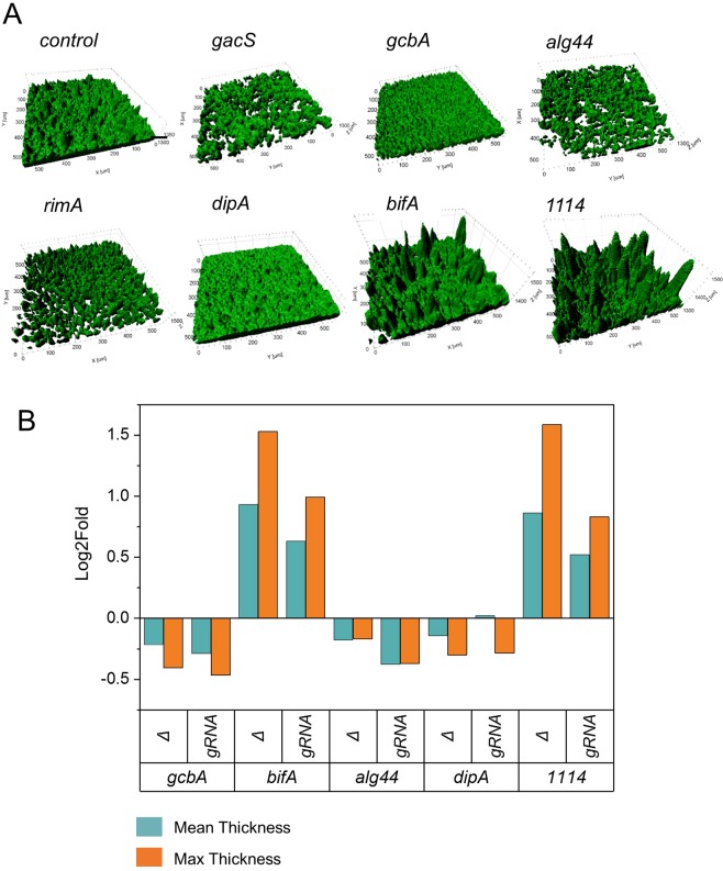
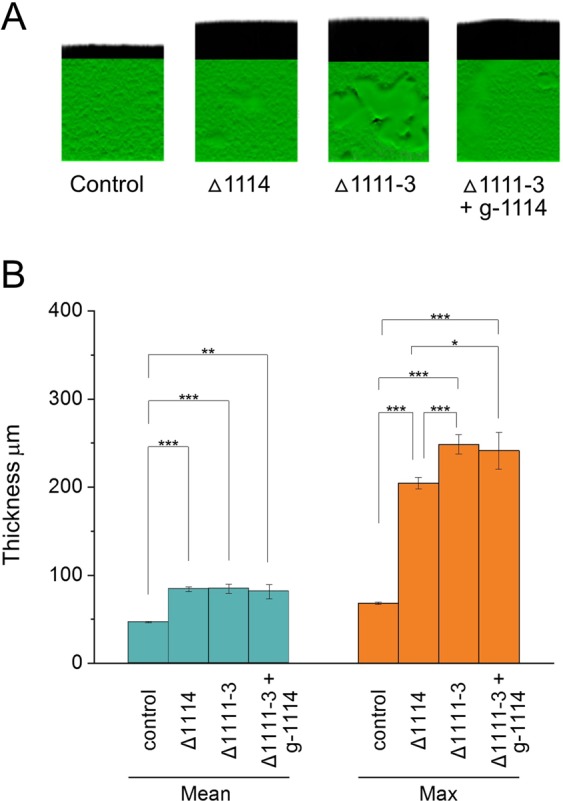
Bacterial biofilm formation involves signaling and regulatory pathways that control the transition from motile to sessile lifestyle, production of extracellular polymeric matrix, and maturation of the biofilm 3D structure. Biofilms are extensively studied because of their importance in biomedical, ecological and industrial settings. Gene inactivation is a powerful approach for functional studies but it is often labor intensive, limiting systematic gene surveys to the most tractable bacterial hosts. Here, we adapted the CRISPR interference (CRISPRi) system for use in diverse strain isolates of P. fluorescens, SBW25, WH6 and Pf0-1. We found that CRISPRi is applicable to study complex phenotypes such as cell morphology, motility and biofilm formation over extended periods of time. In SBW25, CRISPRi-mediated silencing of genes encoding the GacA/S two-component system and regulatory proteins associated with the cylic di-GMP signaling messenger produced swarming and biofilm phenotypes similar to those obtained after gene inactivation. Combined with detailed confocal microscopy of biofilms, our study also revealed novel phenotypes associated with extracellular matrix biosynthesis as well as the potent inhibition of SBW25 biofilm formation mediated by the PFLU1114 operon. We conclude that CRISPRi is a reliable and scalable approach to investigate gene networks in the diverse P. fluorescens group.


Conflict of interest statement

The authors declare no competing interests.

Figures

Figure 1
Figure 1
Gene silencing by the CRISPRi system in P. fluorescens. SBW25 (A) and WH6 (B) strains contain an identical DNA cassette coding for the mNG fluorescent protein expressed from a constitutive promoter (Pc) inserted at a similar chromosomal locus. Both strains harbor the pPFL-Cas9 plasmid and the pPFL-sgRNA plasmid expressing the gRNA Pc2 targeting transcription elongation of the mNG gene at a site overlapping the start of the ORF (see also Fig. S2). Green fluorescence intensities were monitored by flow cytometry over 7 hours after induction of dCas9 (illustrated by colors, right panels). Upon silencing, fluorescence intensity decreases overtime due to dilution from cell divisions (left panels). Dotted lines represent the expected dilution of the GFP in SBW25 (doubling time 135 min) and WH6 (doubling time 120 min).
Figure 2
Figure 2
CRISPRi silencing of genes involved in cell division and cell morphology in P. fluorescens. Cells harboring the pPFL-dCas9 plasmid and the pPFL-gRNA plasmid expressing a gRNANT targeting the start of the ORFs to block transcription elongation. Control with no guide (left panel); silencing of ftsZ (PFLU0952) using a ftsZNT guide (center panel), and silencing of mreB (PFLU0863) using a mreBNT guide (right panel). Strains were grown 5 hours in the presence or absence of inducer (aTc 0.1 μg/ml). Cells were stained with the membrane fluorescent dye FM 4–64 prior to observation by epifluorescence microscopy. Scale bars indicate 2 μm.
Figure 3
Figure 3
Swarming motility phenotypes in P. fluorescens SBW25. Swarming phenotypes were compared for CRISPRi-mediated silencing and deletion mutants. CRISPRi silencing used gRNANT targeting the start of the ORF (denoted g-gene name). Target genes include gacS (PFLU3777) encoding the kinase sensor protein GacS and several genes encoding c-di-GMP binding proteins such as RimA (PFLU0263, PDE), DipA (PFLU0458, PDE), the alginate co-polymerase Alg44 (PFLU0988), GcbA (PFL0621, DGC) and BifA (PFLU4858, PDE) (see also Supplementary Fig. S6). The control corresponds to the pPFL-gRNA plasmid with no guide RNA inserted. Note that SBW25 cells deleted for the same genes also carried the non-active CRISPRi system (i.e., no gRNA expressed) to treat all the strains under identical conditions. (A) Typical swarming morphotypes observed at the surface of soft-agar plates incubated 48 hrs at 25 °C. Plates are circled in purple for CRISPRi silencing (gRNA) and in brown for deletion mutants (Δ). (B) Box plot representing the swarm areas of cells with silenced (gRNA) or deleted (Δ) gene relative to the control strain (n ≥ 4). Statistical significance is indicated (T test, *p <  = 0.05 **p <  = 0.01 ***p <  = 0.001).
Figure 4
Figure 4
CRISPRi-mediated phenotypes associated with biofilm structure and EPS matrix. P. fluorescens SBW25 cells expressing the CRISPRi system targeting genes gacS (PFLU3777, two-component sensor kinase), rimA (PFLU0263, PDE), dipA (PFLU0458, PDE), alg44 (PFLU0988, alginase co-polymerase Alg44), gcbA (PFL0621, DGC) and bifA (PFLU4858, PDE). The control corresponds to the pPFL-gRNA plasmid with no guide RNA inserted. (A) Cells were stained with FM 1–43 and observed by confocal microscopy. 3D architecture of biofilms were reconstructed as described in Methods. Virtual shadow projections were included on the right to show thickness. (B) Biofilms grown in the presence of the Congo Red dye to reveal the structure of EPS. The ESP-stained volumes were rendered with maximum intensity projection. Inset shows a blow-up view of a clump in the EPS matrix after silencing of bifA.
Figure 5
Figure 5
Biofilm phenotypes of genes involved in GacS and c-di-GMP signaling are consistent for CRISPRi silencing and deletion. (A) Surface rendering (IMARIS software) of reconstituted 3D confocal volume images of biofilm pellicles displayed in Fig. 4. Targeted genes encode the GacS (PFLU3777) kinase sensor and the c-di-GMP binding proteins RimA (PFLU0263, PDE), DipA (PFLU0458, PDE), Alg44 (PFLU0988) alginate co-polymerase, GcbA (PFL0621, DGC) and BifA (PFLU4858, PDE). The control corresponds to the pPFL-gRNA plasmid with no guide RNA inserted. (B) Comparison of changes in biofilm thickness caused by CRISPRi silencing and gene deletion.
Figure 6
Figure 6
Role of the PFLU1114 operon in biofilm mass and structure. Quantification of the 3D reconstructed volumes in air-liquid biofilm pellicles (see Fig. 4) were performed using IMARIS x64 9.0.2 XTension software package. (A) Pseudo-3-D images with shadow projection. (B) Histograms displaying the distribution of data obtained from observation of biofilm pellicles (n ≥ 6) from two independent experiments. Pairwise comparisons were performed using the Tukey Method (*p <= 0.05 **p <= 0.01 ***p <= 0.001).

References

    1. Flemming HC, et al. Biofilms: an emergent form of bacterial life. Nature reviews. Microbiology. 2016;14:563–575. doi: 10.1038/nrmicro.2016.94. - DOI - PubMed
    1. Hobley L, Harkins C, MacPhee CE, Stanley-Wall NR. Giving structure to the biofilm matrix: an overview of individual strategies and emerging common themes. FEMS microbiology reviews. 2015;39:649–669. doi: 10.1093/femsre/fuv015. - DOI - PMC - PubMed
    1. Flemming HC, Wingender J. The biofilm matrix. Nature reviews. Microbiology. 2010;8:623–633. doi: 10.1038/nrmicro2415. - DOI - PubMed
    1. McDougald D, Rice SA, Barraud N, Steinberg PD, Kjelleberg S. Should we stay or should we go: mechanisms and ecological consequences for biofilm dispersal. Nature reviews. Microbiology. 2011;10:39–50. doi: 10.1038/nrmicro2695. - DOI - PubMed
    1. Balcazar JL, Subirats J, Borrego CM. The role of biofilms as environmental reservoirs of antibiotic resistance. Frontiers in microbiology. 2015;6:1216. doi: 10.3389/fmicb.2015.01216. - DOI - PMC - PubMed

Publication types

MeSH terms