Review
. 2020 Feb;20(2):74-88.
doi: 10.1038/s41568-019-0216-7. Epub 2019 Nov 4.

The PI3K-AKT network at the interface of oncogenic signalling and cancer metabolism

Affiliations
Review

The PI3K-AKT network at the interface of oncogenic signalling and cancer metabolism

Gerta Hoxhaj et al. Nat Rev Cancer. 2020 Feb.

Abstract

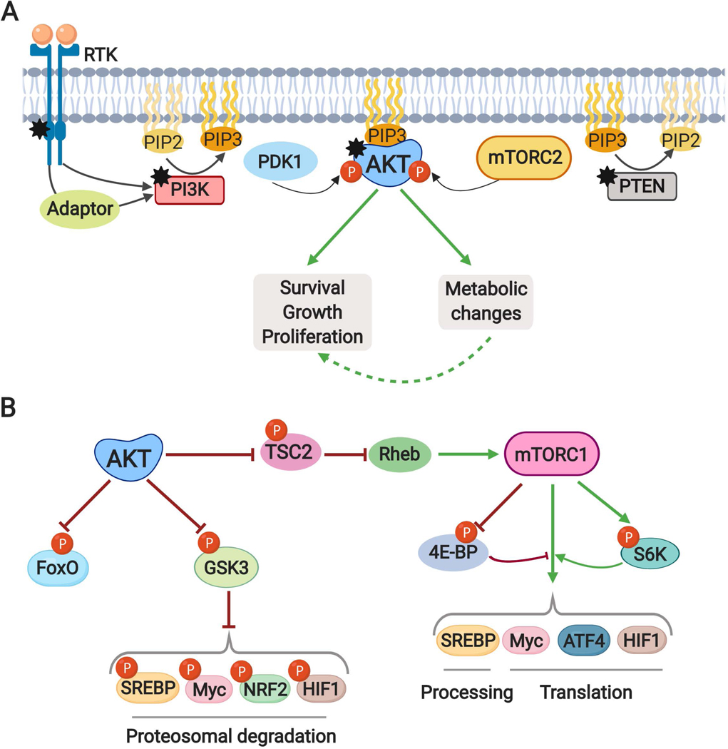
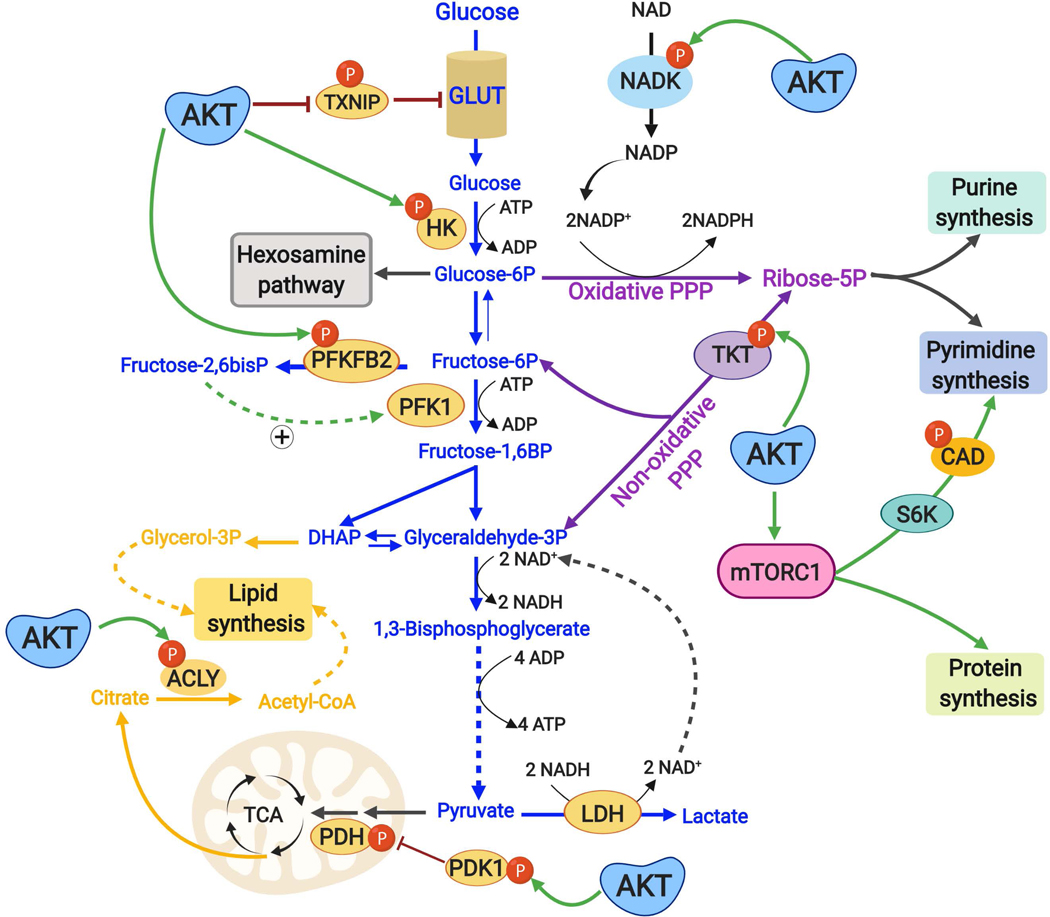
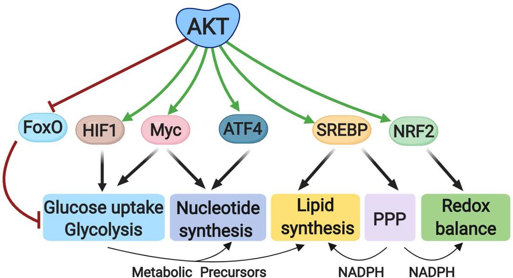
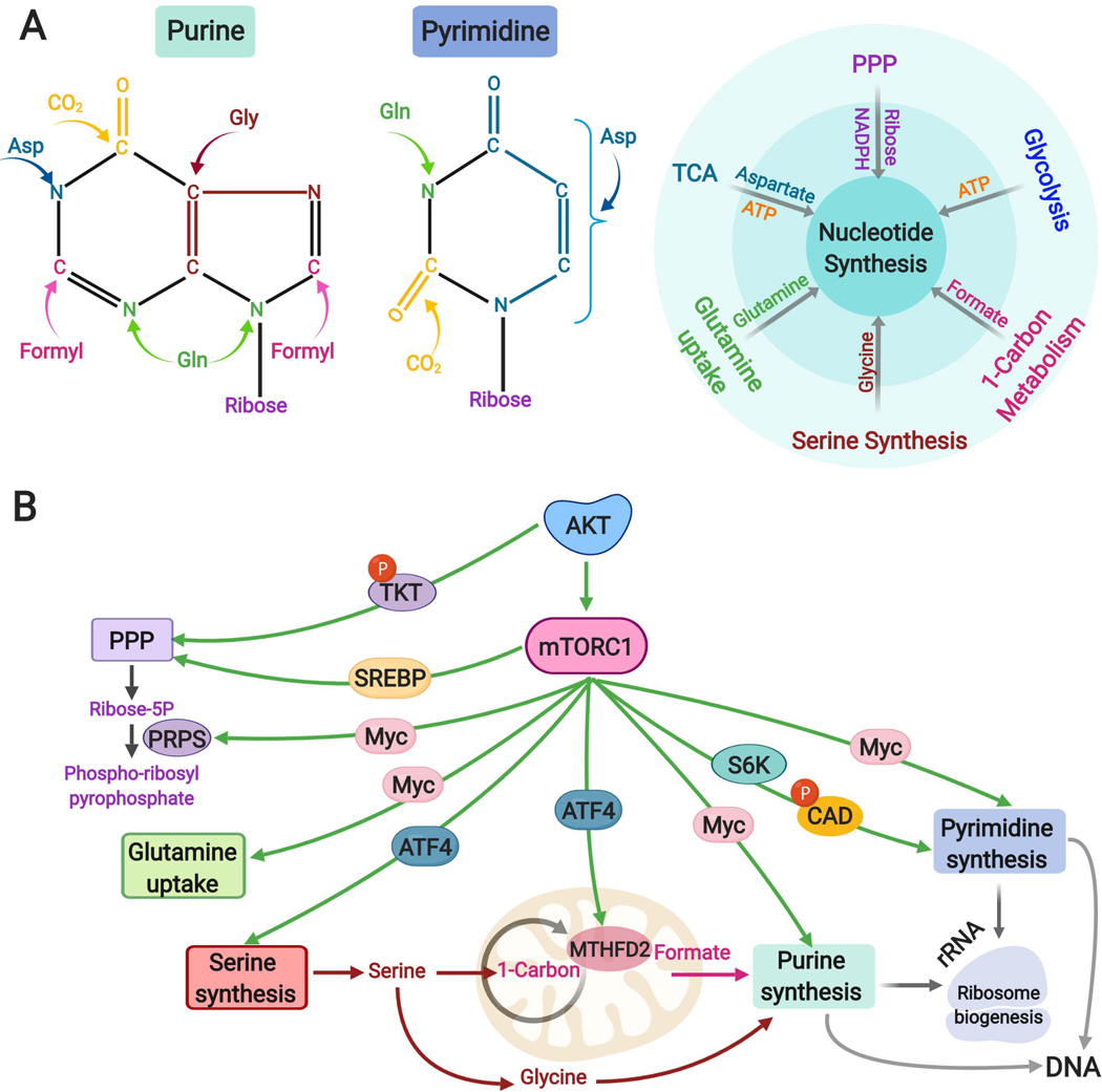
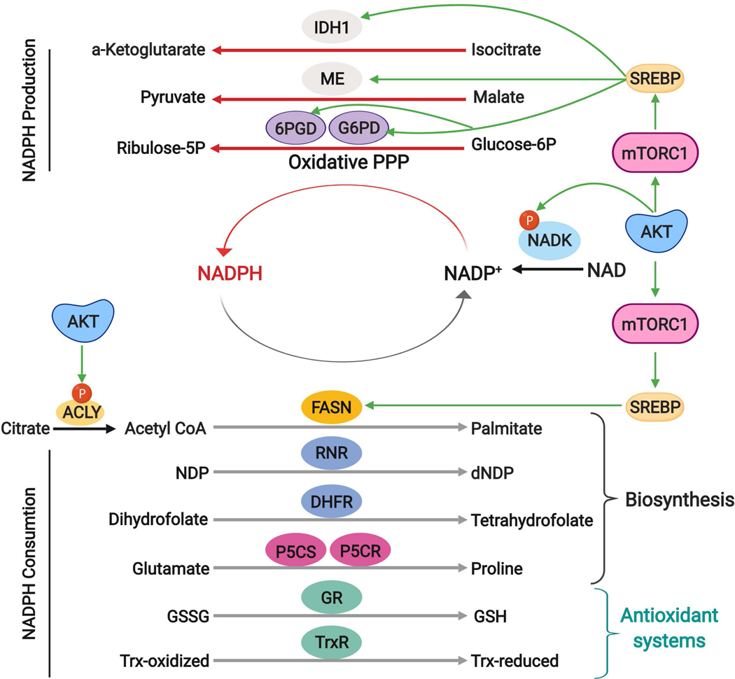
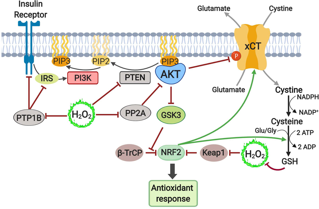
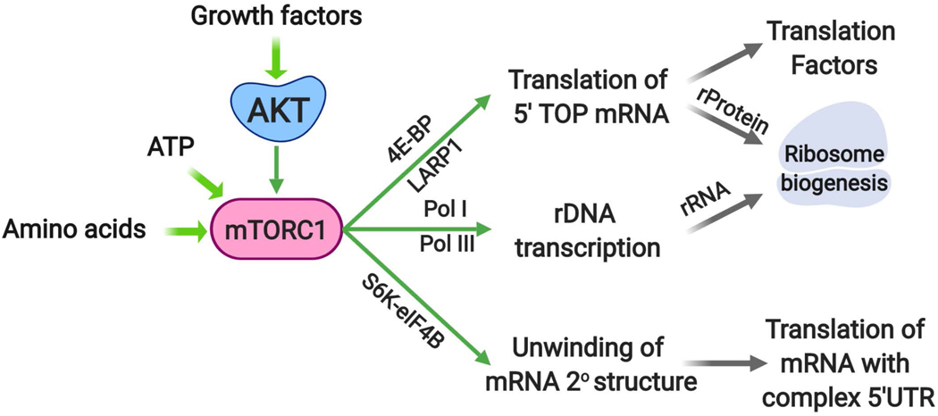
The altered metabolic programme of cancer cells facilitates their cell-autonomous proliferation and survival. In normal cells, signal transduction pathways control core cellular functions, including metabolism, to couple the signals from exogenous growth factors, cytokines or hormones to adaptive changes in cell physiology. The ubiquitous, growth factor-regulated phosphoinositide 3-kinase (PI3K)-AKT signalling network has diverse downstream effects on cellular metabolism, through either direct regulation of nutrient transporters and metabolic enzymes or the control of transcription factors that regulate the expression of key components of metabolic pathways. Aberrant activation of this signalling network is one of the most frequent events in human cancer and serves to disconnect the control of cell growth, survival and metabolism from exogenous growth stimuli. Here we discuss our current understanding of the molecular events controlling cellular metabolism downstream of PI3K and AKT and of how these events couple two major hallmarks of cancer: growth factor independence through oncogenic signalling and metabolic reprogramming to support cell survival and proliferation.


Figures

Fig. 1:
Fig. 1:. The PI3K-AKT pathway and its major downstream effectors.
A. Mechanisms of AKT activation. Receptor tyrosine kinase (RTK) activation and tyrosine phosphorylation of its cytosolic domain or of scaffolding adaptors create binding sites that recruit the lipid kinase PI3K to the plasma membrane. PI3K phosphorylates phosphatidylinositol 4,5-bisphosphate (PIP2) to produce phosphatidylinositol 3,4,5-trisphosphate (PIP3), which can be dephosphorylated back to PIP2 by the lipid phosphatase PTEN. PIP3 acts as a second messenger to recruit the serine/threonine protein kinase AKT to the plasma membrane, where it is fully activated through phosphorylation at T308 and S473 by the PDK1 and mTORC2 protein kinases, respectively. AKT signaling serves to promote cell survival, growth, and proliferation, in part, by inducing various changes to cellular metabolism. Coloured hexagons denote common points of activation and inactivation by cancer-associated mutations. “P” indicates protein phosphorylation events. B. AKT controls cellular metabolism, in part, through three key downstream substrates: TSC2, GSK3, and the FOXO transcription factors. AKT phosphorylates and inhibits TSC2, a component of the TSC complex, to activate mTORC1 by relieving TSC complex-mediated inhibition of Rheb. S6K1 and 4EBP1 are canonical downstream targets of mTORC1, which together with other targets, serve to stimulate the processing and activation of the SREBP family of transcription factors and the mRNA translation of the MYC, HIF1α, and ATF4 transcription factors. GSK3-mediated phosphorylation of the transcription factors SREBP, MYC, NRF2, and HIF1α targets them for ubiquitination and proteasomal degradation. PI3K, phosphoinositide 3-kinase; PTEN, phosphatase and tensin homolog; PDK1, phosphoinositide-dependent protein kinase 1; mTORC2, mechanistic target of rapamycin (mTOR) complex 2; mTORC1, mTOR complex 1; TSC2; tuberous sclerosis complex 2; GSK3, Glycogen synthase kinase 3; FOXO, forkhead box O; Rheb, Ras homolog enriched in brain; S6K1, Ribosomal protein S6 kinase 1; 4EBP1, eukaryotic translation initiation factor 4E binding protein, SREBP: Sterol regulatory element binding proteins; HIF1, Hypoxia-inducible factor 1, ATF4, activating transcription factor 4, NRF2, nuclear factor erythroid 2-related factor 2.
Fig. 2:
Fig. 2:. Direct post-translational regulation of metabolic enzymes and processes downstream of the PI3K-AKT pathway.
AKT stimulates metabolic changes that contribute to anabolic metabolism by directly phosphorylating key metabolic enzymes. AKT promotes plasma membrane localization of the glucose transporter GLUT1 and increased glucose uptake by directly phosphorylating and inhibiting TXNIP, a protein that promotes endocytosis of GLUT1. AKT signaling also promotes retention and metabolic activation of the newly acquired glucose by activating HK2, which phosphorylates glucose to generate glucose 6-phosphate, which cannot be transported out of the cell by GLUT1 and is the entry metabolite for the hexosamine pathway, the oxidative pentose phosphate pathway (PPP), and glycolysis. AKT enhances flux into glycolysis through phosphorylation and activation of PFKFB2, which produces fructose-2,6-bisphosphate, an allosteric activator of the rate-limiting glycolytic enzyme PFK1, which commits the glucose-derived carbon to glycolysis. Both the oxidative and non-oxidative PPP, which branch off of glycolytic intermediates, generate ribose-5-phosphate that serves as the sugar moiety for purine and pyrimidine nucleotides. AKT phosphorylates and activates the non-oxidative PPP enzyme TKT, thereby contributing to ribose-5-phosphate production for nucleotides. AKT also phosphorylates and increases the activity of NADK, which catalyses the phosphorylation of NAD+ to generate NADP+, a limiting substrate for the oxidative PPP, which generates two molecules of the reducing cofactor NADPH through its oxidation of glucose 6-phosphate. Downstream of AKT, mTORC1 activation acutely stimulates de novo pyrimidine synthesis through an S6K-dependent phosphorylation of the pyrimidine synthesis enzyme CAD. Pyruvate, the end product of glycolysis, is either converted to lactate by LDH, which regenerates NAD+ needed for sustained glycolysis, or can enter the mitochondria and TCA cycle for oxidation initiated by the pyruvate dehydrogenase (PDH) complex, the activity of which can be inhibited by pyruvate dehydrogenase kinase (PDK). AKT phosphorylates PDK and promotes its inhibition of PDH, thus favouring the LDH reaction [Note: this phosphorylation is believed to occur within the mitochondria]. AKT directly regulates lipid synthesis through phosphorylation of ACLY, which generates acetyl-CoA in cytosol from the TCA cycle-derived citrate. TXNIP, thioredoxin-interacting protein; GLUT1, glucose transporter 1; PPP, Pentose Phosphate Pathway; PFKFB2, 6-phosphofructo-2-kinase/fructose-2,6-biphosphatase; PFK1, phosphofructokinae 1; TKT, transketolase; NADK, NAD kinase; mTORC1, mTOR complex 1; CAD, carbamoyl-phosphate synthetase 2, aspartate transcarbamylase, and dihydroorotase, S6K1, ribosomal protein S6 kinase 1; NAD+, nicotinamide adenine dinucleotide, NADP+, NAD+ phosphate, LDH, lactate dehydrogenase.
Fig. 3:
Fig. 3:. Transcriptional control of metabolic processes downstream of AKT signaling.
AKT regulates metabolism through a number of downstream transcription factors that control the expression of genes encoding metabolic enzymes. FOXO, HIF1 and MYC regulate the expression of glucose transporters and glycolytic enzymes, and intermediates of glycolysis contribute to nucleotide and lipid synthesis. MYC and ATF4 induce the expression of enzymes contributing to nucleotide synthesis. SREBP isoforms globally induce the expression of lipogenic enzymes, as well as enzymes in the oxidative PPP, which can provide NADPH as reducing equivalents for lipid synthesis and redox. NRF2 regulates redox homeostasis. FOXO, forkhead box O; HIF1, Hypoxia-inducible factor 1; ATF4, activating transcription factor 4, SREBP: Sterol regulatory element binding proteins; NRF2, nuclear factor erythroid 2-related factor 2 and NADPH, nicotinamide adenine dinucleotide phosphate (reduced).
Fig. 4:
Fig. 4:. Regulation of nucleotide metabolism downstream of the AKT-mTORC1 pathway.
A. Left to right: Schematic of purine and pyrimidine nucleotides indicating the donors of carbon and nitrogen atoms that form nucleotides. The small molecules are color coded according to the contributing metabolic pathways from which they are commonly derived, shown schematically to the right. B. Transcriptional and post-translational mechanisms contributing to de novo nucleotide synthesis downstream of the AKT-mTORC1 pathway. Left to right: AKT-mediated phosphorylation of the non-oxidative PPP enzyme TKT and SREBP-mediated regulation of the oxidative PPP enhance the production of ribose-5-phosphate, which can then be used for nucleotide synthesis by conversion to phospho-ribosyl pyrophosphate through the enzyme PRPS, the levels of which are elevated upon MYC activation. MYC promotes glutamine uptake and stimulates expression of several genes encoding enzymes of both purine and pyrimidine synthesis pathways. In addition to being downstream of mTORC1, SREBP and MYC can also be stabilized via AKT-mediated inhibition of GSK3. ATF4 activation downstream of mTORC1 contributes to enhanced purine synthesis through the induction of serine biosynthesis enzymes and the mitochondrial tetrahydrofolate cycle enzyme MTHFD2, thus supplying both glycine and one-carbon formyl units. mTORC1 acutely stimulates pyrimidine synthesis through the S6K1-mediated phosphorylation of the pyrimidine synthesis enzyme CAD. The newly synthesized purine and pyrimidines are used for the synthesis of RNA, predominantly rRNA for ribosome biogenesis, and DNA in proliferating cells. NADPH, nicotinamide adenine dinucleotide phosphate (reduced), PPP, Pentose Phosphate Pathway; TKT, transketolase, SREBP: Sterol regulatory element binding proteins; PRPS, phosphoribosyl pyrophosphate synthase; MTHFD2, methylenetetrahydrofolate dehydrogenase 2, ATF4, activating transcription factor 4; mTORC1, mTOR complex 1.
Fig. 5:
Fig. 5:. AKT signaling and control of NADPH production and consumption.
NADPH serves as a major electron donor for reductive biosynthesis and defence against ROS, reactions that yield its oxidized form, NADP+. In the cytosol, NADPH can be regenerated from NADP+ through reactions involving two enzymes of the oxidative PPP (G6PD and PGD), isocitrate dehydrogenase 1 (IDH1) and malic enzyme (ME). Downstream of AKT and mTORC1, SREBP induces expression of these enzymes to enhance NADPH production. AKT-mediated phosphorylation of NADK serves to boost NADP+ abundance to further enhance the production of NADPH through these redox reactions. Many cellular reactions consume a large quantity of NADPH as reducing power. NADPH is required for key reactions in the biosynthesis of macromolecules, including fatty acid synthase (FASN) to produce palmitate, ribonucleotide reductase (RNR) to convert ribonucleotide diphosphates (NDPs) into deoxyribonucleotide diphosphates (dNDPs), dihydrofolate reductase (DHFR) for tetrahydrofolate synthesis, and the proline synthesis enzymes pyrroline-5-carboxylate (P5C) synthase (P5CS) and pyrroline-5-carboxylate reductase (P5CR). NADPH also serves as essential reducing power for antioxidant enzymes including glutathione reductase (GR), which reduces the oxidized glutathione disulfide (GSSG) to glutathione (GSH), and thioredoxin reductase (TrxR), which transfers electrons from NADPH to reduce thioredoxin for subsequent reduction of oxidized cysteines. ROS, reactive oxygen species; PPP, pentose phosphate pathway; NADPH, nicotinamide adenine dinucleotide phosphate (reduced); G6PD, glucose 6-phosphate dehydrogenase; PGD, phosphogluconate dehydrogenase; SREBP: Sterol regulatory element binding proteins; NADK, NAD kinase.
Figure 6:
Figure 6:. Interplay between ROS and the PI3K-AKT pathway
ROS in the form of hydrogen peroxide can activate the PI3K-AKT pathway through inactivation of protein phosphatases, including PTP1B, which attenuates the activity of insulin receptor and insulin receptor substrate (IRS), and PP2A, which normally dephosphorylates T308 on AKT, thus leading to enhanced AKT phosphorylation and activation in response to ROS. ROS also inhibits the lipid phosphatase PTEN, resulting in accumulation of PIP3 and activation of AKT. AKT activation can facilitate the adaptation to ROS by activating the NRF2 transcription factor, which stimulates numerous enzymes to mount an antioxidant response. Downstream of AKT, GSK3 phosphorylates NRF2 and marks it for ubiquitination by the β-TRCP E3 ligase, leading to subsequent proteasomal degradation. Thus, through its inhibition of GSK3, AKT signaling can stabilize NRF2. NRF2 is stabilized and activated by ROS via cysteine oxidation of the E3 ligase Keap1, thereby disrupting its binding of NRF2. AKT also directly phosphorylates and inhibits the cystine-glutamate antiporter xCT, which transports cystine into cells that, upon NADPH-dependent reduction to cysteine, can be used to produce glutathione for ROS neutralization. xCT and the enzymes of glutathione synthesis are encoded by gene targets of NRF2 as part of its anti-oxidant response. ROS, Reactive oxygen species; PTP1B, protein tyrosine phosphatase 1B; PP2A, protein phosphatase 2A; PTEN, phosphatase and tensin homolog; PDK1, phosphoinositide-dependent protein kinase 1; NRF2, nuclear factor erythroid 2-related factor 2; GSK3, Glycogen synthase kinase 3; NADPH, nicotinamide adenine dinucleotide phosphate (reduced), Keap1, kelch-Like ECH-associated protein 1; β-TRCP, β-transducin repeat containing protein.
None

References

    1. Lawrence MS et al. Discovery and saturation analysis of cancer genes across 21 tumour types. Nature 505, 495–501 (2014). - PMC - PubMed
    1. Fruman DA et al. The PI3K pathway in human disease. Cell 170, 605–635 (2017). - PMC - PubMed
    1. Thorpe LM, Yuzugullu H & Zhao JJ PI3K in cancer: divergent roles of isoforms, modes of activation and therapeutic targeting. Nat Rev Cancer 15, 7–24 (2015). - PMC - PubMed
    1. Alessi DR et al. Mechanism of activation of protein kinase B by insulin and IGF-1. EMBO J 15, 6541–6551 (1996). - PMC - PubMed
    1. Alessi DR et al. Characterization of a 3-phosphoinositide-dependent protein kinase which phosphorylates and activates protein kinase Balpha. Curr Biol 7, 261–269 (1997). - PubMed

Publication types