. 2020 Jan;16(1):7-14.
doi: 10.1038/s41589-019-0378-3. Epub 2019 Nov 4.

Structural complementarity facilitates E7820-mediated degradation of RBM39 by DCAF15

Affiliations

Structural complementarity facilitates E7820-mediated degradation of RBM39 by DCAF15

Tyler B Faust et al. Nat Chem Biol. 2020 Jan.

Abstract

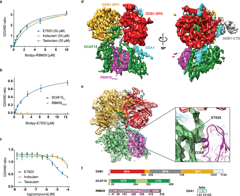
The investigational drugs E7820, indisulam and tasisulam (aryl-sulfonamides) promote the degradation of the splicing factor RBM39 in a proteasome-dependent mechanism. While the activity critically depends on the cullin RING ligase substrate receptor DCAF15, the molecular details remain elusive. Here we present the cryo-EM structure of the DDB1-DCAF15-DDA1 core ligase complex bound to RBM39 and E7820 at a resolution of 4.4 Å, together with crystal structures of engineered subcomplexes. We show that DCAF15 adopts a new fold stabilized by DDA1, and that extensive protein-protein contacts between the ligase and substrate mitigate low affinity interactions between aryl-sulfonamides and DCAF15. Our data demonstrate how aryl-sulfonamides neo-functionalize a shallow, non-conserved pocket on DCAF15 to selectively bind and degrade RBM39 and the closely related splicing factor RBM23 without the requirement for a high-affinity ligand, which has broad implications for the de novo discovery of molecular glue degraders.


Conflict of interest statement

Competing interests: E.S.F. is a founder and/or member of the scientific advisory board (SAB), and equity holder of C4 Therapeutics and Civetta Therapeutics and a consultant to Novartis, AbbVie and Pfizer. The Fischer lab receives research funding from Novartis, Deerfield and Astellas. N.S.G. is a founder, SAB member and equity holder in Gatekeeper, Syros, Petra, C4, B2S and Soltego. The Gray lab receives or has received research funding from Novartis, Takeda, Astellas, Taiho, Janssen, Kinogen, Voronoi, Her2llc, Deerfield and Sanofi. N.S.G., E.S.F, H.Y., Q.C., T.Z., T.F., R.P.N and K.A.D. are inventors on a patent application (PCT/US2018/065701 and PCT/US2019/014919), submitted by the Dana-Farber Cancer Institute.

Figures

Fig. 1 |
Fig. 1 |. Cryo-EM structure of the DDB1ΔB-DCAF15-DDA1 complex bound to E7820 and RBM39RRM2.
a, TR-FRET. Titration of BodipyFL-RBM39RRM2 to DDB1ΔB-DCAF15biotin in the presence of E7820 (1, KDapp = 2.0 μM), indisulam (2, KDapp = 2.1 μM), or tasisulam (3, KDapp = 3.5 μM) at 50 μM. b, TR-FRET. Titration of BodipyFL-E7820 (4) probe to DDB1ΔB-DCAF15biotin or RBM39RRM2-biotin. Compound binding is only observed for DDB1ΔB-DCAF15biotin (KDapp = 3.8 μM). c, Competitive titration of BodipyFL-E7820 (4) with aryl sulfonamides in TR-FRET assay. DDB1ΔB-DCAF15biotin is at 200 nM, BodipyFL-E7820 (4) is at 5 μM, and aryl-sulfonamides are at 0.002–100 μM. TR-FRET data in a-c are plotted as means ± s.d. from three independent replicates (n = 3). d, 4.4 Å cryo-EM map of the DDB1ΔB-DCAF15-DDA1-E7820-RBM39RRM2 complex segmented to indicate DDA1 (cyan), DCAF15 (green), RBM39RRM2 (magenta), DDB1-BPC (orange), DDB1-BPA (red), and DDB1-CTD (grey). e, Cryo-EM map shown with the fitted and refined model. (Right), close-up of the region of the RBM39-DCAF15 interface, with the resistance mutation site G268V indicated in yellow and the putative E7820 density outlined in dotted lines. f, Domain representation of the proteins present in the complex. Regions omitted from the constructs are indicated by hatched lines.
Fig. 2 |
Fig. 2 |. Crystal structure of the DDB1ΔB-DCAF15split-DDA1-E7820-RBM39RRM2 complex.
a, (Left) Cartoon representation of the DDB1ΔB-DCAF15-DDA1-E7820-RBM39RRM2 complex. DDA1 (cyan), DCAF15-NTD (blue), DCAF15-CTD (green), RBM39RRM2 (magenta), DDB1-BPC (orange), DDB1-BPA (red), and DDB1-CTD (grey). E7820 is shown as spheres. (Right) A different view of the complex, shown in transparent surface representation. b, Cartoon representation of DCAF15 indicating secondary structure elements and colored in blue and green, for the DCAF15-NTD and DCAF15-CTD, respectively. DCAF15 alpha helices and beta strands are numbered from the N- to C-terminus, which are shown as colored circles for both the NTD and CTD of DCAF15. c, Cartoon view of DCAF15, highlighting the five stacked β-sheets. Helices from the NTD and CTD are colored in grey.
Fig. 3 |
Fig. 3 |. DDA1 stabilizes the CRL4DCAF15 complex and facilitates RBM39 recruitment.
a, Cartoon representation of the DDB1ΔB-DCAF15split-E7820-RBM39 complex with DDA1 highlighted as a cyan surface representation. DDA1 binds at the top of DDB1-BPA, winds down the back side of the propeller, and ends in a helix buried in DCAF15. b, DDB1 and DCAF15 are shown as a grey and green surface, respectively, and DDA1 is represented as a cartoon colored according to the conservation scores as calculated in ConSurf. The top 3 bins of conservation in ConSurf (high conservation) are colored in red, orange, and yellow, respectively, while the bottom 6 bins (average and variable conservation, shown as “low”) are colored in gray to highlight the most conserved surfaces. c, TR-FRET. Titration of BodipyFL-RBM39RRM2 to DDB1ΔB-DCAF15biotin (KDapp = 1.9 μM) or DDB1ΔB-DCAF15biotin-DDA1 (KDapp = 0.62 μM) in the presence of E7820 (50 μM), demonstrating enhanced recruitment of RBM39RRM2 to the DDA1-containing complex. d, TR-FRET. Titration of E7820 to DDB1ΔB-DCAF15biotin (EC50 = 0.74 μM) or DDB1ΔB-DCAF15biotin-DDA1 and BodipyFL-RBM39RRM2 (EC50 = 0.33 μM). e, Titration of BodipyFL-E7820 to DDB1ΔB-DCAF15biotin (KDapp = 3.8 μM) or DDB1ΔB-DCAF15biotin-DDA1 (KDapp = 3.8 μM). TR-FRET data in c-e are plotted as means ± s.d. from three independent replicates (n = 3).
Fig. 4 |
Fig. 4 |. Aryl-sulfonamide binding to DCAF15.
a, Sketch of E7820 and its interactions with DCAF15 and RBM39. Water-mediated hydrogen bonds are highlighted in cyan. b, Chemical structures of E7820 (1), indisulam (2), and tasisulam (3). c, E7820 interacts predominantly through the sulfonamide moiety and the indole moiety with residues in the DCAF15-NTD (blue). Additional hydrophobic interactions with the DCAF15-CTD (green), and sulfur-π interaction as well as water (cyan)-mediated hydrogen bonds with RBM39 (magenta) stabilize E7820 in a shallow pocket. d, Surface representation of DCAF15 is shown in grey and E7820, indisulam and tasisulam are shown as stick representation in yellow, magenta and cyan, respectively.
Fig. 5 |
Fig. 5 |. Inter-protein contacts between DCAF15 and RBM39.
a, Surface representation of DCAF15 and RBM39RRM2 indicating the extensive interacting interface on DCAF15 and RBM39, shown in grey. E7820 is shown as a yellow stick representation. b, Side chain interactions between DCAF15, RBM39 and E7820. RBM39 buries a large hydrophobic surface on the DCAF15 α7 helix, in addition to four salt-bridges with DCAF15 on the opposing side of the binding interface. c, Scatter plot depicting identification of the novel E7820 substrate, RBM23, in Kelly cells. Kelly cells were treated with E7820 (10 μM) for 5 hours, and protein abundance was analyzed using TMT quantification mass spectrometry (two-sided moderated t-test as implemented in limma, n = 3 for dmso, n = 1 for E7820). d, Alignment of the second RRM domain from RBM39 and RBM23. Residues in black are completely conserved, gray shading represents similar substitutions, and white indicates no conservation. Red circles above the alignment indicate the positions of resistance mutations in RBM39 for indisulam-dependent toxicity. e, TR-FRET. Titration of E7820 to DDB1ΔB-DCAF15 in the presence of BodipyFL-RBM39RRM2-WT (EC50 = 0.74 μM), BodipyFL-RBM23RRM2-WT (EC50 = 1.0 μM). TR-FRET data in e are plotted as means ± s.d. from three independent replicates (n = 3).
Fig. 6 |
Fig. 6 |. Topological and evolutionary constraints on E7820 activity.
a, A model of the CRL4DCAF15 ligase bound to E7820 and RBM39RRM2. The N- and C-termini of RBM39RRM2 (pink circles) are positioned near RBX1 in the ligase, while RBM39RRM2 itself is bound on a non-proximal side face of DCAF15. The DCAF15split crystal structure was superimposed onto the DDB1-DDB2-CUL4A-RBX1 crystal structure (pdb: 4a0k). b, Evolutionary conservation of DCAF15 (top) and CRBN (bottom). The substrate receptors are represented as a surface, colored according to the conservation scores as calculated in ConSurf with the top 3 bins of conservation colored in red, orange, and yellow, respectively, and the bottom 6 bins colored in gray to highlight the most conserved surfaces. DCAF15 is shown bound to E7820 (yellow) and the α1 helix (residues 262–274) of RBM39RRM2 (magenta), while CRBN is shown bound to lenalidomide (green) and the β-hairpin loop (residues 29–49) of CK1α (cyan). Lenalidomide and CK1α both bind in a highly conserved pocket of CRBN.

Comment in

  • Molecular glue concept solidifies.
    Baek K, Schulman BA. Baek K, et al. Nat Chem Biol. 2020 Jan;16(1):2-3. doi: 10.1038/s41589-019-0414-3. Nat Chem Biol. 2020. PMID: 31819271 No abstract available.

References

    1. Salami J & Crews CM Waste disposal-An attractive strategy for cancer therapy. Science 355, 1163–1167, doi:10.1126/science.aam7340 (2017). - DOI - PubMed
    1. Chamberlain PP et al. Structure of the human Cereblon-DDB1-lenalidomide complex reveals basis for responsiveness to thalidomide analogs. Nat Struct Mol Biol 21, 803–809 (2014). - PubMed
    1. Fischer ES, Park E, Eck MJ & Thoma NH SPLINTS: small-molecule protein ligand interface stabilizers. Curr Opin Struct Biol 37, 115–122, doi:10.1016/j.sbi.2016.01.004 (2016). - DOI - PMC - PubMed
    1. Fischer ES et al. Structure of the DDB1-CRBN E3 ubiquitin ligase in complex with thalidomide. Nature 512, 49–53 (2014). - PMC - PubMed
    1. Ito T et al. Identification of a primary target of thalidomide teratogenicity. Science (New York, NY) 327, 1345–1350 (2010). - PubMed

Methods-only References

    1. Abdulrahman W et al. A set of baculovirus transfer vectors for screening of affinity tags and parallel expression strategies. Anal Biochem 385, 383–385 (2009). - PubMed
    1. Kabsch W Xds. Acta Crystallogr D Biol Crystallogr 66, 125–132, doi:10.1107/S0907444909047337 (2010). - DOI - PMC - PubMed
    1. Winn MD et al. Overview of the CCP4 suite and current developments. Acta crystallographica. Section D, Biological crystallography 67, 235–242, doi:10.1107/S0907444910045749 (2011). - DOI - PMC - PubMed
    1. Mccoy A et al. Phaser crystallographic software. Journal of Applied Crystallography 40, 658–674 (2007). - PMC - PubMed
    1. Emsley P & Cowtan K Coot: model-building tools for molecular graphics. Acta Crystallographica Section D-Biological Crystallography 60, 2126–2132 (2004). - PubMed

Publication types

MeSH terms

Substances