Case Reports
. 2019 Nov;25(11):1680-1683.
doi: 10.1038/s41591-019-0611-3. Epub 2019 Nov 4.

Resistance to autosomal dominant Alzheimer's disease in an APOE3 Christchurch homozygote: a case report

Affiliations
Case Reports

Resistance to autosomal dominant Alzheimer's disease in an APOE3 Christchurch homozygote: a case report

Joseph F Arboleda-Velasquez et al. Nat Med. 2019 Nov.

Abstract

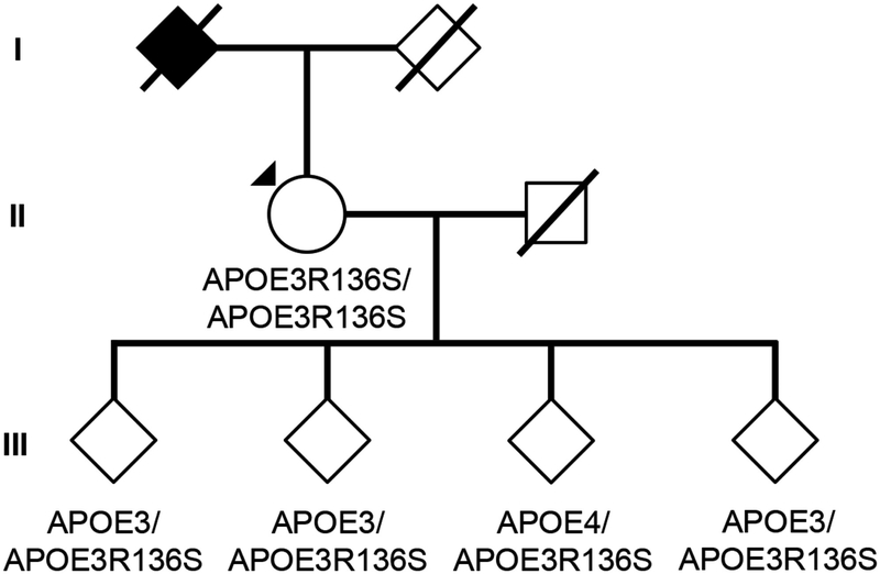
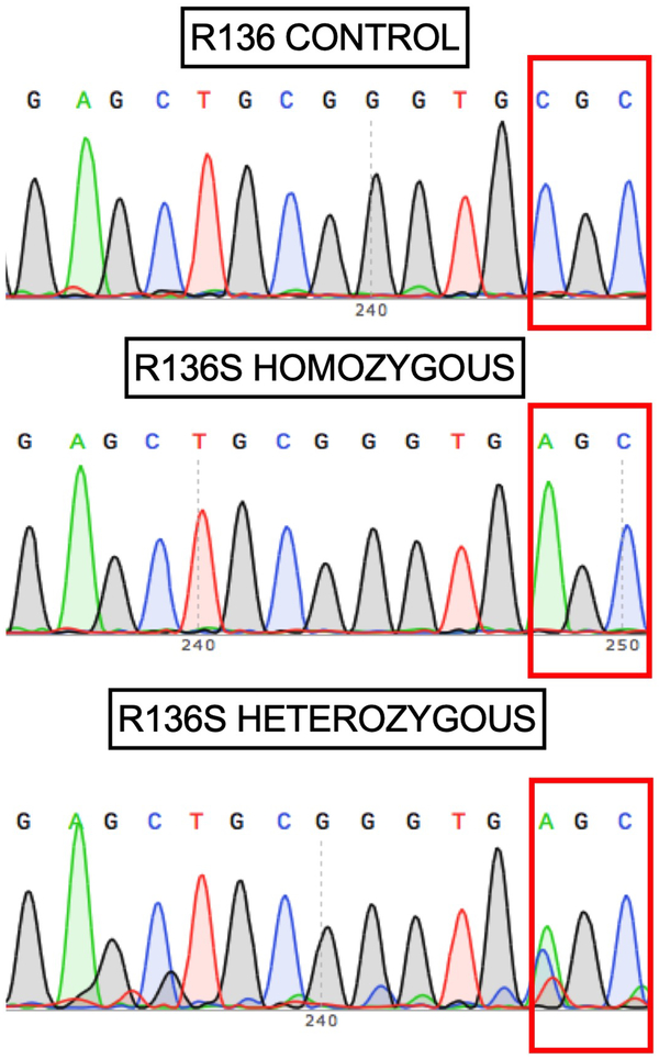
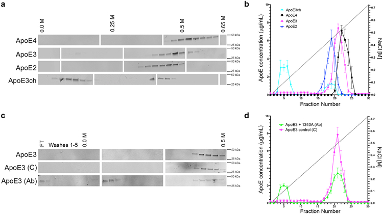
We identified a PSEN1 (presenilin 1) mutation carrier from the world's largest autosomal dominant Alzheimer's disease kindred, who did not develop mild cognitive impairment until her seventies, three decades after the expected age of clinical onset. The individual had two copies of the APOE3 Christchurch (R136S) mutation, unusually high brain amyloid levels and limited tau and neurodegenerative measurements. Our findings have implications for the role of APOE in the pathogenesis, treatment and prevention of Alzheimer's disease.


Conflict of interest statement

Competing interests

P.N.T. has received personal compensation for consulting, serving on scientific advisory boards, or other activities with AbbVie, AC Immune, Acadia, Auspex, Boehringer Ingelheim, Chase Pharmaceuticals, Corium, Eisai, GliaCure, INSYS Therapeutics, Pfizer, T3D, AstraZeneca, Avanir, Biogen, Eli Lilly, H. Lundbeck A/S, Merck and Company, and Roche; holds a provisional patent on “Biomarkers of Alzheimer’s Disease” at the University of Rochester; holds stock options in Adamas; received research support from AstraZeneca, Avanir, Biogen, Eli Lilly, H. Lundbeck A/S, Merck and Company, Roche, Amgen, Avid, Functional Neuromodulation, GE Healthcare, Genentech, Novartis, Takeda, Targacept, the National Institute on Aging, and the Arizona Department of Health Services. H.Z. has served at scientific advisory boards for Roche Diagnostics, Wave, Samumed and CogRx, has given lectures in symposia sponsored by Biogen and Alzecure, and is a co-founder of Brain Biomarker Solutions in Gothenburg AB, a GU Ventures-based platform company at the University of Gothenburg (outside submitted work). R.A.S. has received personal compensation from AC Immune, Eisai, Roche, and Takeda, and research grant support from Eli Lilly and Janssen. All other authors declare no competing financial interests.

Figures

Fig. 1
Fig. 1. Subject’s genealogy
Circles represent females, squares represent males, diamonds represent individuals whose gender has been masked for privacy, arrowhead depicts proband individual with MCI, and shading indicates individual with history of dementia. Deceased individuals are marked with a crossed bar. APOE genotypes are indicated as appropriate to preserve anonymity. Family links were verified by three family informants. Genotypes of relatives of the R136S homozygote individual were determined by Sanger sequencing (Extended Data Figure 2).
Fig. 2
Fig. 2. Sanger DNA sequencing of homozygous ApoEch carrier individual
Representative direct sequencing results of amplicon in APOE gene from control, proband, and descendants’ samples. R136 homozygous sequence is shown in upper panel from a control individual. In the middle panel, proband case is shown. R136S homozygous mutation can be observed. In bottom panel a R136S heterozygous mutation from a descendant is identified. Data representative of n=2 independent experiments and further validated independently with whole exome and whole genome sequencing.
Fig. 3
Fig. 3. ApoE3ch modulates Aβ aggregation
(A) Rate of Aβ42 fibril formation in the presence of C-terminus fragments of ApoE3 wild-type (WT), APOE3ch, or in the absence of ApoE as detected by Thioflavin T fluorescence. Changes in relative fluorescence units (RFU) were plotted for time in minutes (min). Aβ42 fibrillation rate was lower in the presence of ApoE3ch compared to ApoE3 WT (****p = 0.00000797 for 55 μM Aβ42 + ApoE3WT vs. 55 μM Aβ42 + ApoE3ch; ****p = 0.0000000042 for 55 μM Aβ42 vs. 55 μM Aβ42 + ApoE3WT; ***p = 0.00022 for 55 μM Aβ42 vs. 55 μM Aβ42 + ApoE3ch, 2-way ANOVA followed by Tukey’s multiple comparisons test; n=3 represended as mean ± SEM.). (B) Schematic representation of the split-luciferase complementation triggered by amyloid oligomerization in vitro. (C) Percentage of luminescence expressed as relative luminescence units (RLU) was obtained by split-luciferase complementation assay after 24 hours in culture media from 293T cells transfected with full-length ApoE3ch or ApoE3WT cDNA. Luciferase luminescence by oligomer formation is significantly reduced in ApoE3ch compared to ApoE3 WT indicating lower aggregation. (*p = 0.0202, 2-tailed unpaired T-test, n=6). Representative data presented as individual values ± SEM of n=3 independent experiments.
Figure 1.
Figure 1.. Brain imaging shows limited tau pathology and neurodegeneration despite high amyloid-β plaque burden in an individual homozygous for APOE3ch.
A, PET measurements of amyloid plaque burden (top row, PiB DVRs) and PHF tau (bottom row, flortaucipir standard uptake value ratios (FTP SUVRs). PET images are superimposed onto the medial and lateral surfaces of the left hemisphere. Color-coded scale bar indicates PiB DVR or FTP SUVR values; blue represents lowest binding and red represents highest binding, ranging from 0.8 to 2.0. The APOE3ch homozygote (left panels) had greater amyloid-β plaque burden and relatively limited PHF tau burden, particularly for her age, compared to PSEN1 E280A mutation carriers with MCI at the kindred’s typical age of 44 years. In line with institutional review board regulations, PET imaging measurements were not repeated within short time intervals. B, 18F-fludeoxyglucose (FDG) PET precuneus cerebral metabolic rate for glucose (CMRgl) images are shown. Color-coded scale bar indicates FDG SUVR values; blue represents lowest values and red represents highest values, ranging of 0.5 to 2.1. The arrow and circle indicate that the individual homozygous for APOE3ch had relatively preserved CMRgl in a precuneus/posterior cingulate region known to be preferentially affected by Alzheimer’s disease. ChC, Christchurch. C, Brain imaging measurements of mean cortical amyloid plaque burden, inferior temporal cortex PHF tau burden, hippocampal volume, and precuneus glucose metabolism in the PSEN1 E280A mutation carrier with two APOE3ch alleles and exceptionally late onset of MCI (red dots), PSEN1 mutation carriers with MCI onset at the kindred’s typical, younger age (black dots) and PSEN1 mutation carriers who have not yet developed MCI (gray dots). Amyloid plaque burden is expressed as mean cortical-to-cerebellar distribution volume ratios (DVRs). Paired helical filament (PHF) tau burden is expressed as inferior temporal-to-cerebellar FTP SUVRs. Hippocampal volumes are expressed as hippocampal-to-whole brain volume ratios. Cerebral glucose metabolism is reflected as precuneus-to-whole-brain CMRgl ratios. While the PSEN1 E280A mutation carrier with two APOE3ch alleles had by far the highest amyloid plaque burden, she did not have comparably severe PHF tau burden or hippocampal atrophy, and she had no evidence of precuneus glucose hypometabolism.
Figure 2.
Figure 2.. The Christchurch mutation impairs ApoE’s heparin binding.
(A, C) We used western blotting to detect ApoE in protein fractions eluted from heparin columns using an increasing NaCl gradient. Individual blots were cropped between 25 to 50 kDa. Blank spaces separate individual blots that are representative of n = 2 independent experiments. FT = flow through. (B, D) We used ELISA to quantify differences in the NaCl elution pattern of different ApoE isoforms from heparin columns. N = 3 columns per isoform in independent experiments were analyzed side-by-side twice on different days to quantify differences. Error bars depict standard error of mean. In (B) ApoE2 is depicted in blue; ApoE3, magenta; ApoE4, black; and, ApoE3ch, cyan. In (D) wild type ApoE3 in the presence of an ApoE-specific antibody (1343A) is depicted in green whereas control is shown in magenta. ApoE3ch was eluted from the heparin column with the lowest NaCl concentration revealing impaired heparin binding compared to other ApoE isoforms (A, C). Wild type ApoE3 was also eluted with low NaCl concentrations when incubated with an antibody specific for an HSPG-binding domain of ApoE (B, D).

References

    1. Quiroz YT, et al. Association Between Amyloid and Tau Accumulation in Young Adults With Autosomal Dominant Alzheimer Disease. JAMA Neurol 75, 548–556 (2018). - PMC - PubMed
    1. Llado A, et al. A novel PSEN1 mutation (K239N) associated with Alzheimer’s disease with wide range age of onset and slow progression. Eur J Neurol 17, 994–996 (2010). - PubMed
    1. Thordardottir S, et al. Reduced penetrance of the PSEN1 H163Y autosomal dominant Alzheimer mutation: a 22-year follow-up study. Alzheimers Res Ther 10, 45 (2018). - PMC - PubMed
    1. Knight WD, et al. Pure progressive amnesia and the APPV717G mutation. Alzheimer Dis Assoc Disord 23, 410–414 (2009). - PubMed
    1. Velez JI, et al. APOE*E2 allele delays age of onset in PSEN1 E280A Alzheimer’s disease. Mol Psychiatry 21, 916–924 (2016). - PMC - PubMed

Publication types

MeSH terms