

Molecular Mechanisms and Therapeutics for SBMA/Kennedy's Disease
- PMID: 31686397
- PMCID: PMC6985201
- DOI: 10.1007/s13311-019-00790-9
Molecular Mechanisms and Therapeutics for SBMA/Kennedy's Disease
Abstract
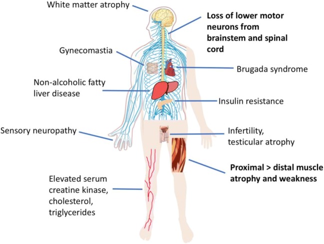
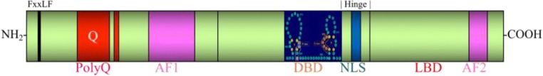
Spinal and bulbar muscular atrophy (SBMA) is a neuromuscular disease caused by a polyglutamine (polyQ) expansion in the androgen receptor (AR). Despite the fact that the monogenic cause of SBMA has been known for nearly 3 decades, there is no effective treatment for this disease, underscoring the complexity of the pathogenic mechanisms that lead to a loss of motor neurons and muscle in SBMA patients. In the current review, we provide an overview of the system-wide clinical features of SBMA, summarize the structure and function of the AR, discuss both gain-of-function and loss-of-function mechanisms of toxicity caused by polyQ-expanded AR, and describe the cell and animal models utilized in the study of SBMA. Additionally, we summarize previously conducted clinical trials which, despite being based on positive results from preclinical studies, proved to be largely ineffective in the treatment of SBMA; nonetheless, these studies provide important insights as researchers develop the next generation of therapies.
Keywords: Polyglutamine; androgen receptor; motor neuron; neurodegenerative disease; spinal and bulbar muscular atrophy..
Figures
References
-
- Kennedy WR, Alter M, Sung JH. Progressive proximal spinal and bulbar muscular atrophy of late onset. A sex-linked recessive trait. Neurology. 1968;18(7):671–80. - PubMed
-
- La Spada AR, Wilson EM, Lubahn DB, Harding AE, Fischbeck KH. Androgen receptor gene mutations in X-linked spinal and bulbar muscular atrophy. Nature. 1991;352(6330):77–79. - PubMed
-
- Atsuta N, Watanabe H, Ito M, Banno H, Suzuki K, Katsuno M, et al. Natural history of spinal and bulbar muscular atrophy (SBMA): a study of 223 Japanese patients. Brain. 2006;129(Pt 6):1446–55. - PubMed
-
- Sobue G, Hashizume Y, Mukai E, Hirayama M, Mitsuma T, Takahashi A. X-linked recessive bulbospinal neuronopathy. A clinicopathological study. Brain. 1989;112(Pt 1):209–32. - PubMed
Publication types
MeSH terms
Substances
LinkOut - more resources
Full Text Sources
Other Literature Sources
Research Materials
