. 2019 Dec 3;58(48):4882-4892.
doi: 10.1021/acs.biochem.9b00705. Epub 2019 Nov 13.

Helix N-Cap Residues Drive the Acid Unfolding That Is Essential in the Action of the Toxin Colicin A

Affiliations

Helix N-Cap Residues Drive the Acid Unfolding That Is Essential in the Action of the Toxin Colicin A

Yan Huang et al. Biochemistry. .

Abstract

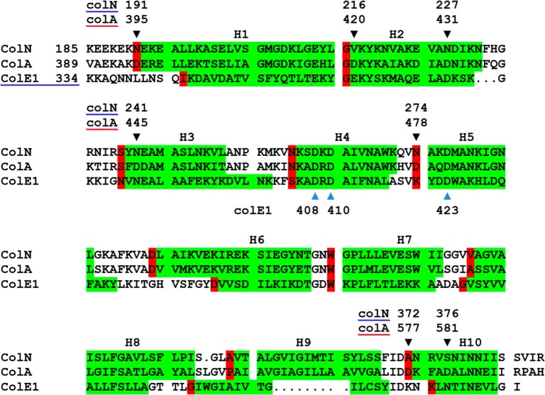
Numerous bacterial toxins and other virulence factors use low pH as a trigger to convert from water-soluble to membrane-inserted states. In the case of colicins, the pore-forming domain of colicin A (ColA-P) has been shown both to undergo a clear acidic unfolding transition and to require acidic lipids in the cytoplasmic membrane, whereas its close homologue colicin N shows neither behavior. Compared to that of ColN-P, the ColA-P primary structure reveals the replacement of several uncharged residues with aspartyl residues, which upon replacement with alanine induce an unfolded state at neutral pH. Here we investigate ColA-P's structural requirement for these critical aspartyl residues that are largely situated at the N-termini of α helices. As previously shown in model peptides, the charged carboxylate side chain can act as a stabilizing helix N-Cap group by interacting with free amide hydrogen bond donors. Because this could explain ColA-P destabilization when the aspartyl residues are protonated or replaced with alanyl residues, we test the hypothesis by inserting asparagine, glutamine, and glutamate residues at these sites. We combine urea (fluorescence and circular dichroism) and thermal (circular dichroism and differential scanning calorimetry) denaturation experiments with 1H-15N heteronuclear single-quantum coherence nuclear magnetic resonance spectroscopy of ColA-P at different pH values to provide a comprehensive description of the unfolding process and confirm the N-Cap hypothesis. Furthermore, we reveal that, in urea, the single domain ColA-P unfolds in two steps; low pH destabilizes the first step and stabilizes the second.


Conflict of interest statement

The authors declare no competing financial interest.

Figures

Figure 1
Figure 1
Sequence alignment of colicin pore-forming domains. C-Terminal pore-forming domains of colicin N (UniProtKB P08083), colicin A (UniProtKB P04480), and colicin E1 (UniProtKB P02978) are shown. Helices are colored green, and N-Cap positions red. Residues mutated in this study are shown by black triangles (▼) with numbering for full length colicin A and N. Residues thought to be involved in acid destabilization of colicin E1 are shown with blue triangles.
Figure 2
Figure 2
Colicin A P-domain WT unfolding characteristics. (A) Urea-dependent unfolding of ColA-P at acidic pH measured by the shift of the barycentric mean wavelength (BMW) of the intrinsic tryptophan fluorescence. Samples were equilibrated in 50 mM citrate buffer and urea for >8 h before measurement or 50 mM phosphate for pH 7.0. (B) Effect of temperature on the far-UV and (inset) near-UV CD spectrum of ColA-P at pH 7.0. Solid line for 25 °C and dashed line for 80 °C. (C) Thermal transitions for near-UV CD measured at 295 nm (left ordinate axis) and far-UV CD measured at 222 nm (right ordinate axis). (D) DSC scans (1 °C/min) of ColA-P solutions at different pH values.
Figure 3
Figure 3
Alanine substitution mutants mainly destabilize the first urea unfolding transition. (A) Urea denaturation of ColA-P Asp to Ala mutants at pH 7.0. (B) GdnHCl denaturation of Asp to Ala mutants at pH 7.0. (C) Urea denaturation of the ColA-P D431A mutant at low pH. (D) Urea denaturation of the ColA-P D577A mutant at low pH. BMW is the barycentric mean wavelength.
Figure 4
Figure 4
Thermal denaturation data reflect the urea denaturation results. Panels A and B show circular dichroism. The intensity was measured at (A) 222 nm to measure secondary structure (α helix) content and (B) 295 nm to measure the tertiary structure signal provided by buried aromatic residues. (C) DSC thermograms for each mutant at pH 7.0.
Figure 5
Figure 5
Asp to Asn mutant data. (A) Unfolding of mutants induced by urea and measured by intrinsic tryptophan fluorescence at pH 7.0. BMW is the barycentric mean wavelength of intrinsic tryptophan fluorescence. (B) GdnHCl unfolding measured in the same way. (C) DSC thermograms.
Figure 6
Figure 6
Glutamate and glutamine insertions at three N-Cap sites show intermediate thermal stability. Differential scanning calorimetry data (1 °C/min) for each of the ColA-P mutants in 50 mM phosphate buffer (pH 7.0). For reference, D420E has wild type stability.
Figure 7
Figure 7
N-Cap glutamate and glutamine insertions are more stabilizing than predicted in urea denaturation experiments. Effects of glutamine and glutamate replacement mutants at three N-Cap positions on urea-induced unfolding at pH 7.0 and 3.0. Unfolding is followed by the change in the barycentric mean wavelength (BMW) of the intrinsic tryptophan fluorescence. Results are also shown for the relevant asparagine and alanine mutations and the WT (see Figures 2–5). Panels A, C, and E show pH 7.0 results for mutations at positions D420, D478, and D577, respectively, and panels B, D, and F show pH 3.0 results for mutations at positions D420, D478, and D577, respectively.
Figure 8
Figure 8
NMR shows that D420A retains its low-pH conformation at pH 8.0. 1H–15N HSQC spectra of 15N-labeled ColA-P. The region of data mainly shows peptide backbone amide (N–H) resonances. (A) The wild type ColA-P 1H spectrum shows a general shift to a higher field when the pH is increased to 8.0. (B) WT and D420A spectra at pH 4.5 show similar distributions. (C) The D420A spectrum at pH 8.0 remains similar to the WT spectrum at pH 4.5. (D) As expected from the information presented above, at pH 8.0, WT and D420A spectra show different distributions. The mutant thus retains its pH 4.5 spectrum at pH 8.0. The NMRpeak positions are available in the Supporting Information.

References

    1. Abrami L.; Liu S.; Cosson P.; Leppla S. H.; van der Goot F. G. (2003) Anthrax toxin triggers endocytosis of its receptor via a lipid raft-mediated clathrin-dependent process. J. Cell Biol. 160, 321–328. 10.1083/jcb.200211018. - DOI - PMC - PubMed
    1. Chenal A.; Prongidi-Fix L.; Perier A.; Aisenbrey C.; Vernier G.; Lambotte S.; Fragneto G.; Bechinger B.; Gillet D.; Forge V.; Ferrand M. (2009) Deciphering Membrane Insertion of the Diphtheria Toxin T Domain by Specular Neutron Reflectometry and Solid-State NMR Spectroscopy. J. Mol. Biol. 391, 872–883. 10.1016/j.jmb.2009.06.061. - DOI - PubMed
    1. Rodnin M. V.; Li J.; Gross M. L.; Ladokhin A. S. (2016) The pH-Dependent Trigger in Diphtheria Toxin T Domain Comes with a Safety Latch. Biophys. J. 111, 1946–1953. 10.1016/j.bpj.2016.09.030. - DOI - PMC - PubMed
    1. Sandvig K.; Olsnes S. (1980) Diphtheria-toxin entry into cells is facilitated by low pH. J. Cell Biol. 87, 828–832. 10.1083/jcb.87.3.828. - DOI - PMC - PubMed
    1. Bullough P. A.; Hughson F. M.; Skehel J. J.; Wiley D. C. (1994) Structure of influenza hemagluttinin at the pH of membrane fusion. Nature 371, 37–43. 10.1038/371037a0. - DOI - PubMed

Publication types