

Paired-agent imaging for detection of head and neck cancers
- PMID: 31686720
- PMCID: PMC6827556
- DOI: 10.1117/12.2510897
Paired-agent imaging for detection of head and neck cancers
Abstract
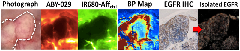
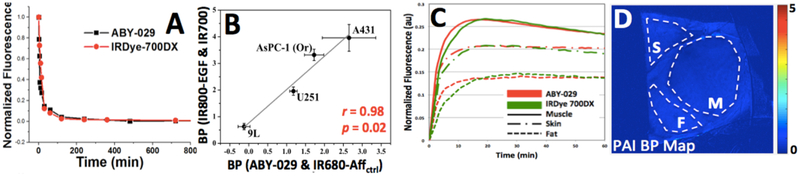
Head and neck cancers overwhelmingly overexpress epidermal growth factor receptor (EGFR). This overexpression has been utilized for head and neck cancers using molecular targeted agents for therapy and cancer cell detection. Significant progress has been made in using EGFR-targeted fluorescent antibody and Affibody molecule agents for fluorescent guided surgery in head and neck cancers. Although success in achieving tumor-to-background ratio of 3-5 have been achieved, the field is limited by the non-specific fluorescence in normal tissues as well as EGFR specific fluorescence in the oral cavity. We propose that paired-agent imaging (PAI) could improve the contrast between tumor and normal tissue by removing the fluorescent signal arising from non-specific binding. Here, ABY-029 - an anti-EGFR Affibody molecule labeled with IRDye 800CW - and IRDye 680RD conjugated to Affibody Control Imaging Agent molecule (IR680-Affctrl) are used as targeted and untargeted control agents, respectively, in a panel of head and neck squamous cell carcinomas (HNSCC) to test the ability of PAI to increase tumor detection. Initial results demonstrate that binding potential, a value proportional to receptor concentration, correlates well to EGFR expression but experimental limitations prevented pixel-by-pixel analysis that was desired. Although promising, a more rigorous and well-defined experimental protocol is required to align ex vivo EGFR immunohistochemistry with in vivo binding potential and fluorescence intensity. Additionally, a new set of paired-agents, ABY-029 and IRDye 700DX, are successfully tested in naïve mice and will be carried forward for clinical translation.
Keywords: ABY-029; Affibody Control Imaging Agent; Affibody molecule; IRDye 680RD; IRDye 700DX; IRDye 800CW; epidermal growth factor receptor; fluorescence guided surgery; head and neck cancer; paired-agent imaging.
Figures


Similar articles
-
Identification of a Suitable Untargeted Agent for the Clinical Translation of ABY-029 Paired-Agent Imaging in Fluorescence-Guided Surgery.Mol Imaging Biol. 2023 Feb;25(1):97-109. doi: 10.1007/s11307-021-01642-9. Epub 2021 Oct 12. Mol Imaging Biol. 2023. PMID: 34642897 Free PMC article.
-
Preclinical imaging of epidermal growth factor receptor with ABY-029 in soft-tissue sarcoma for fluorescence-guided surgery and tumor detection.J Surg Oncol. 2019 Jun;119(8):1077-1086. doi: 10.1002/jso.25468. Epub 2019 Apr 4. J Surg Oncol. 2019. PMID: 30950072 Free PMC article.
-
Improved Discrimination of Tumors with Low and Heterogeneous EGFR Expression in Fluorescence-Guided Surgery Through Paired-Agent Protocols.Mol Imaging Biol. 2023 Feb;25(1):110-121. doi: 10.1007/s11307-021-01656-3. Epub 2021 Oct 14. Mol Imaging Biol. 2023. PMID: 34651290 Free PMC article.
-
Current challenges and clinical investigations of epidermal growth factor receptor (EGFR)- and ErbB family-targeted agents in the treatment of head and neck squamous cell carcinoma (HNSCC).Cancer Treat Rev. 2014 May;40(4):567-77. doi: 10.1016/j.ctrv.2013.10.002. Epub 2013 Oct 12. Cancer Treat Rev. 2014. PMID: 24216225 Review.
-
Application of Fluorescence-Guided Surgery to Subsurface Cancers Requiring Wide Local Excision: Literature Review and Novel Developments Toward Indirect Visualization.Cancer Control. 2018 Jan-Mar;25(1):1073274817752332. doi: 10.1177/1073274817752332. Cancer Control. 2018. PMID: 29334791 Free PMC article. Review.
Cited by
-
Receptor-Targeted Fluorescence-Guided Surgery With Low Molecular Weight Agents.Front Oncol. 2021 Jun 30;11:674083. doi: 10.3389/fonc.2021.674083. eCollection 2021. Front Oncol. 2021. PMID: 34277418 Free PMC article. Review.
-
Fluorescence Lifetime Imaging Enables In Vivo Quantification of PD-L1 Expression and Intertumoral Heterogeneity.Cancer Res. 2025 Feb 1;85(3):618-632. doi: 10.1158/0008-5472.CAN-24-0880. Cancer Res. 2025. PMID: 39514403 Free PMC article.
-
Paired Agent Fluorescence Imaging of Cancer in a Living Mouse Using Preassembled Squaraine Molecular Probes with Emission Wavelengths of 690 and 830 nm.Bioconjug Chem. 2020 Feb 19;31(2):214-223. doi: 10.1021/acs.bioconjchem.9b00750. Epub 2019 Dec 6. Bioconjug Chem. 2020. PMID: 31756298 Free PMC article.
-
Dual-channel pulse-dye densitometry can enable correction of fluorescent targeted and control agent plasma input function differences for quantitative paired-agent molecular imaging: a simulation study.J Biomed Opt. 2025 Apr;30(4):046001. doi: 10.1117/1.JBO.30.4.046001. Epub 2025 Mar 29. J Biomed Opt. 2025. PMID: 40161250 Free PMC article.
-
In vivo quantification of programmed death-ligand-1 expression heterogeneity in tumors using fluorescence lifetime imaging.Res Sq [Preprint]. 2023 Oct 23:rs.3.rs-3222037. doi: 10.21203/rs.3.rs-3222037/v1. Res Sq. 2023. PMID: 37961361 Free PMC article. Preprint.
References
-
- Jacobs C, Goffinet D, Goffinet L et al., “Chemotherapy as a substitute for surgery in the treatment of advanced resectable head and neck cancer,” Cancer, 60(6), 1178–83 (1987). - PubMed
-
- Kowalski LP, Magrin J, Waksman G et al., “Supraomohyoid neck dissection in the treatment of head and neck tumors: survival results in 212 cases,” Archives of Otolaryngology–Head & Neck Surgery, 119(9), 958–963 (1993). - PubMed
-
- Ravasz LA, Slootweg PJ, Hordijk GJ et al., “The status of the resection margin as a prognostic factor in the treatment of head and neck carcinoma,” Journal of Cranio-Maxillofacial Surgery, 19(7), 314–318 (1991). - PubMed
-
- Binahmed A, Nason RW, and Abdoh AA, “The clinical significance of the positive surgical margin in oral cancer,” Oral oncology, 43(8), 780–784 (2007). - PubMed
-
- Pillsbury HC, and Clark M, “A rationale for therapy of the N0 neck,” The Laryngoscope, 107(10), 1294–1315 (1997). - PubMed
Grants and funding
LinkOut - more resources
Full Text Sources
Research Materials
Miscellaneous