. 2019 Sep 6:12:7297-7306.
doi: 10.2147/OTT.S207640. eCollection 2019.

In vitro mitochondrial-targeted antioxidant peptide induces apoptosis in cancer cells

Affiliations

In vitro mitochondrial-targeted antioxidant peptide induces apoptosis in cancer cells

Wei Zhan et al. Onco Targets Ther. .

Abstract

Introduction: Reactive oxygen species (ROS) are major contributors to cancer and involved in numerous tumor proliferation signaling pathways. Mitochondria are the major ROS-producing organelles, and ROS are produced from the synthesis of adenosine triphosphate and cell metabolism.

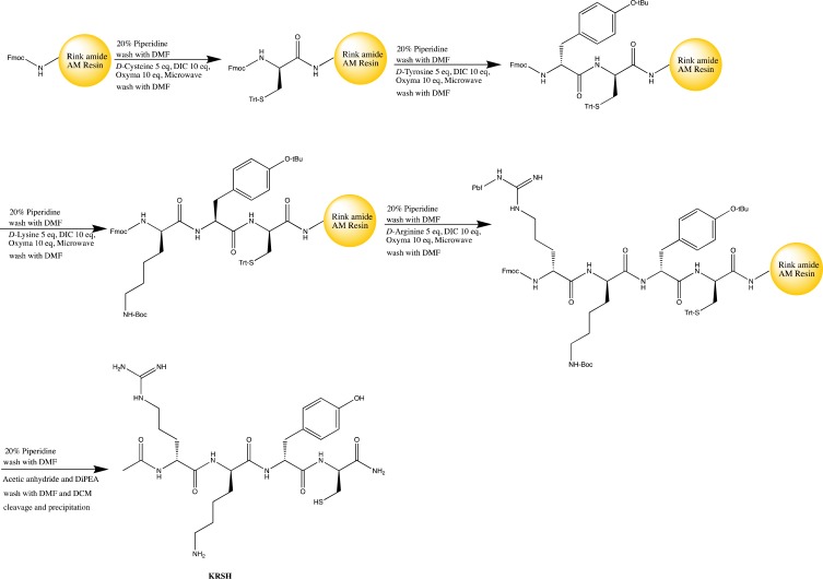
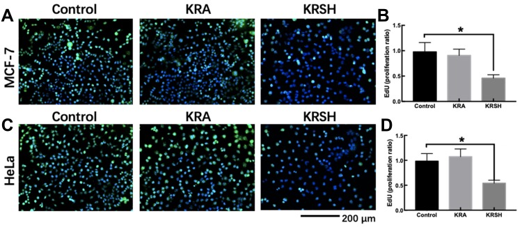
Methods: A novel mitochondria-targeted peptide, namely KRSH, was synthesized and characterized. KRSH consists of four amino acids; lysine and arginine contain positively charged groups that help KRSH target the mitochondria, while tyrosine and cysteine neutralize excessive endogenous ROS, thereby inhibiting tumorigenesis.

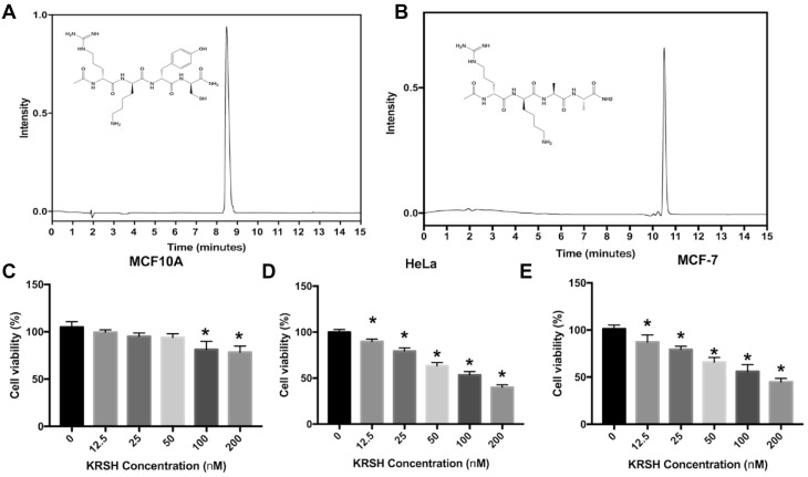
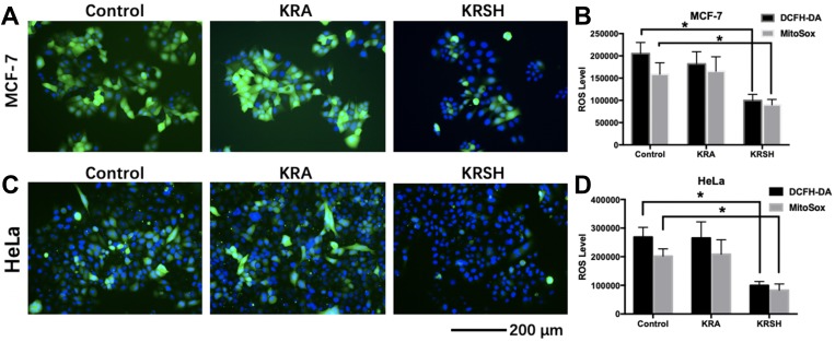
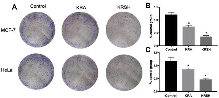
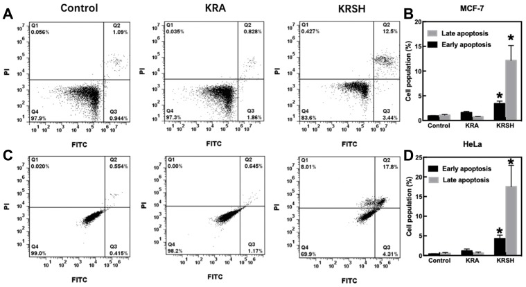
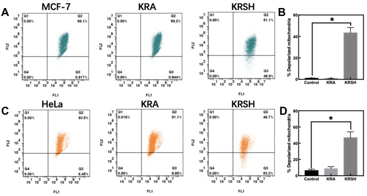
Results: The results indicated that KRSH is specifically inhibiting the growth of HeLa and MCF-7 cancer cell lines. However, MCF10A cells can resist the effects of KRSH even in a relative higher concentration. The dichloro-dihydro-fluorescein diacetate and MitoSOXTM Red assay suggested that KRSH drastically decreased the level of ROS in cancer cells. The mitochondrial depolarization assay indicated that treatment with KRSH at a dose of 50 nM may decrease the mitochondrial membrane potential leading to apoptosis of HeLa and MCF-7 cells.

Conclusion: In other studies, investigating rat liver mitochondria, the uptake of KRSH may reach 80% compared with that for mitoquinone. Therefore, KRSH was designed as a superior peptide antioxidant and a mitochondria-targeting anticancer agent.

Keywords: ROS; anticancer; mitochondrial targeted; oxidative stress; reactive oxygen species.


Conflict of interest statement

The authors report no conflicts of interest in this work.

Figures

Figure 1
Figure 1
The scheme of solid phase peptide synthesis with microwave-aided.
Figure 2
Figure 2
The analytical trace of the KRSH peptide and KRA (negative control) (A, B). Cell viability after treatment with the KRSH peptide at different doses (0–200 nM). The neutral red assay was employed to evaluate the survival rate of MCF10A, HeLa, and MCF-7 cells (C–E). *p<0.05 compared with control cells cultured in complete medium.
Figure 3
Figure 3
The levels of intracellular and mitochondrial ROS in different cell lines after treatment with KRA and the KRSH peptide for 24 h were detected using DCFH-DA and MitoSOXTM, and counterstained with Hoechst stain. The levels of ROS in MCF-7 cells (A, B) and HeLa cells (C, D) decreased significantly after treatment with the KRSH peptide. *p<0.05 compared with control cells cultured in complete medium. Abbreviations: ROS, reactive oxygen species; DCFH-DA, 2′,7′-dichlorofluorescin diacetate.
Figure 4
Figure 4
Colony formation assay was performed on (A, B) HeLa cells and (A, C) MCF-7 cells after treatment with 50 nM KRA and the KRSH peptide for 24 h using the Giemsa stain. The results are expressed as the mean ± SD from at least three independent experiments. *p<0.05 compared with control cells cultured in complete medium. Abbreviation: SD, standard deviation.
Figure 5
Figure 5
EdU staining to detect cell proliferation after treatment with KRSH. (A) MCF-7 cells were treated with EdU for 6 h prior to click reaction, and the data analysis is displayed in the graph (B). The analysis was performed to calculate the signal intensity in EdU-positive cells based on individual DAPI signal. (C) The proliferation of HeLa cells after treatment with the peptide was also detected using the EdU reaction system after a 6-h incubation. The statistical analysis is displayed in the graph (D) using the same calculation. *p<0.05, compared with control cells cultured in complete medium, one-way ANOVA. Abbreviations: EdU, 5-ethynyl-2’-deoxyuridine; DAPI, 4′,6-diamidino-2-phenylindole; ANOVA, analysis of variance.
Figure 6
Figure 6
Cell apoptosis after incubation with 50 nM KRA and the KRSH peptide for 24 h. (A) Apoptosis in MCF-7 cells was distinguished through FITC-Annexin V coupled with PI staining. (B) The corresponding quantification of early and late apoptosis in MCF-7 cells. (C) Apoptosis in HeLa cells was determined using a flow cytometer, and the data analysis is displayed in (D). *p<0.05, compared with control cells cultured in complete medium, one-way ANOVA). Abbreviations: FITC, fluorescein isothiocyanate; ANOVA, analysis of variance.
Figure 7
Figure 7
Mitochondrial membrane depolarization was determined using the JC-1 probe. (A) Depolarization in MCF-7 cells was detected after incubation with the peptide using a flow cytometer. (B) Quantitative analysis of the shift of mitochondrial red fluorescence to green fluorescence among groups, indicating the depolarization of the mitochondrial membrane. (C, D) HeLa cells were examined and analyzed using the same procedure. *p<0.05, compared with control cells cultured in complete medium, one-way ANOVA. Abbreviation: ANOVA, analysis of variance.

References

    1. Weinberg F, Hamanaka R, Wheaton WW, et al. Mitochondrial metabolism and ROS generat ion are essential for Kras-mediated tumorigenicity. Proc Natl Acad Sci U S A. 2010;107(19):8788–8793. doi:10.1073/pnas.1003428107 - DOI - PMC - PubMed
    1. Cai J, Yang J, Jones DP. Mitochondrial control of apoptosis: the role of cytochrome c. Biochim Biophys Acta. 1998;1366(1–2):139–149. doi:10.1016/s0005-2728(98)00109-1 - DOI - PubMed
    1. Donovan M, Cotter TG. Control of mitochondrial integrity by Bcl-2 family members and caspase-independent cell death. Biochim Biophys Acta. 2004;1644(2–3):133–147. doi:10.1016/j.bbamcr.2003.08.011 - DOI - PubMed
    1. Tait SW, Ichim G, Green DR. Die another way–non-apoptotic mechanisms of cell death. J Cell Sci. 2014;127(Pt 10):2135–2144. doi:10.1242/jcs.093575 - DOI - PMC - PubMed
    1. Murphy MP. How mitochondria produce reactive oxygen species. Biochem J. 2009;417(1):1–13. doi:10.1042/BJ20081386 - DOI - PMC - PubMed