. 2019 Oct 7:2019:1892508.
doi: 10.1155/2019/1892508. eCollection 2019.

Cytokine Profile in Early Infection by Leptospira interrogans in A/J Mice

Affiliations

Cytokine Profile in Early Infection by Leptospira interrogans in A/J Mice

Lorena Bavia et al. J Immunol Res. .

Abstract

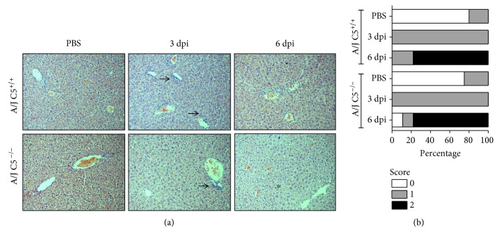
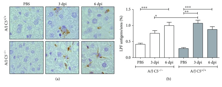
Leptospirosis is considered a neglected disease with an estimated more than one million cases every year. Since rodents are at the same time the main reservoir and generally asymptomatic to Leptospira infection, understanding why some animal species are resistant and others are susceptible to this infection would shed some light in how to control this important zoonosis. The innate immune response against Leptospira is mainly dependent on phagocytosis and activation of the Complement System. In this context, cytokines may drive the early control of infection and the adaptive response. Since the Complement System is important to eliminate leptospires in vivo, we investigated if Complement C5 in A/J mice would modulate the cytokine production during infection by Leptospira interrogans serovar Kennewicki type Pomona Fromm (LPF). Thus, our aim was to investigate the systemic levels of pro- and anti-inflammatory cytokines during Leptospira infection in the blood, liver, lung, and kidney on the third and sixth days of infection in A/J C5+/+ and A/J C5-/- mice. Blood levels of TNF-α, IL-6, IFN-γ, and MCP-1 reached a peak on the third day. Although both mouse strains developed splenomegaly, similar histopathological alterations in the liver and the lung, levels of pro- and anti-inflammatory cytokines were different. A/J C5+/+ mice had higher levels of liver IL-10, IL-1β, IL-12p40, and IL-12p70 and kidney IL-1β, IL-12p40, and IL-12p70 on the sixth day of infection when compared to A/J C5-/- mice. Our results showed that in A/J genetic background, the Complement component C5 modulates a cytokine profile in the liver and kidney of infected mice, which may play a role in the control of disease progression.


Conflict of interest statement

The authors declare that there is no conflict of interest regarding the publication of this paper.

Figures

Figure 1
Figure 1
Liver histopathological analysis in LPF-infected A/J mice. Mice were inoculated i/p with 1.5 × 10⁸ LPF or only PBS and then euthanized on the third or the sixth days postinfection (n ≥ 4). (a) Liver sections (3–5 μm) were stained with HE and evaluated at 200x magnification. Arrows indicate leukocyte infiltration in the portal space and in the hepatic sinusoids. Asterisk indicates mitotic cells. (b) Total scores of hepatic lesions. A/J−/−: C5-deficient mice, PBS (n = 5), 3 dpi (n = 12), 6 dpi (n = 9); A/J C5+/+: congenic C5-sufficient mice PBS (n = 4), 3 dpi (n = 10), 6 dpi (n = 9). dpi: days postinfection.
Figure 2
Figure 2
Lung histopathological analysis in LPF-infected A/J mice. Mice were inoculated i/p with 1.5 × 10⁸ LPF or only PBS and then euthanized on the third or the sixth days postinfection (n ≥ 4). (a) Lung sections (3–5 μm) were stained with HE and evaluated at 200x magnification. The inserts present the immunohistochemical analysis of the lung from LPF-infected mice evaluated at 400x magnification. (b) Total scores of lung lesions. A/J−/−: C5-deficient mice, PBS (n = 5), 3 dpi (n = 12), 6 dpi (n = 9); A/J C5+/+: congenic C5-sufficient mice PBS (n = 4), 3 dpi (n = 10), 6 dpi (n = 9).dpi: days postinfection.
Figure 3
Figure 3
Presence of LPF antigens in the liver of LPF-infected A/J mice. (a) Liver sections from A/J mice were incubated with rabbit antileptospire immune serum. (b) Semiquantitative measurements of LPF antigens in the liver of infected A/J mice were performed in seven random fields of each section. Significant differences are indicated as when p < 0.05, ∗∗p < 0.01, and ∗∗∗p < 0.001. A/J−/−: C5-deficient mice, PBS (n = 5), 3 dpi (n = 12), 6 dpi (n = 9); A/J C5+/+: congenic C5-sufficient mice PBS (n = 4), 3 dpi (n = 10), 6 dpi (n = 9). dpi: days postinfection.
Figure 4
Figure 4
Liver concentrations of pro- and anti-inflammatory cytokines during early infection by LPF. A/J C5+/+ and A/J C5−/− mice were inoculated i/p with 1.5 × 10⁸ LPF or PBS and then euthanized on the third or the sixth days. The concentrations of several cytokines were analyzed in the liver extracts by ELISA, using the same protein concentration in each sample. Significant differences are indicated as when p < 0.05. dpi: days postinfection.
Figure 5
Figure 5
Lung concentrations of pro- and anti-inflammatory cytokines during early infection by LPF. A/J C5+/+ and A/J C5−/− mice were inoculated i/p with 1.5 × 10⁸ LPF or PBS and then euthanized on the third or the sixth days. The concentrations of several cytokines were analyzed in the liver extracts by ELISA, using the same protein concentration in each sample. Significant differences are indicated as when p < 0.05. dpi: days postinfection.
Figure 6
Figure 6
Kidney concentrations of pro- and anti-inflammatory cytokines during early infection by LPF. A/J C5+/+ and A/J C5−/− mice were inoculated i/p with 1.5 × 10⁸ LPF or PBS and then euthanized on the third or the sixth days. The concentrations of several cytokines were analyzed in the liver extracts by ELISA, using the same protein concentration in each sample. Significant differences are indicated as when p < 0.05. dpi: days postinfection.
Figure 7
Figure 7
Cytokine levels are dependent on the presence of Complement component C5 during early infection by LPF. Pro- and anti-inflammatory cytokines were evaluated in the liver, lung, and kidney. The levels of IL-10, IL-1β, and IL-12 (p40 and p70) were higher in the liver of A/J C5+/+ (C5+) versus A/J C5−/− (C5) mice on the sixth day of infection. Although the kidneys are the organs affected in the chronic period of LPF infection, the lack of C5 was important for the production of TNF-α, IL-6, and IL-10 on the third day of infection. However, the presence of C5 promoted the synthesis of IL-1β and IL-12 (p40 and p70). Since no difference was found in lung cytokine levels regarding the presence or lack of C5, this organ was omitted. dpi: days postinfection.

Similar articles

Cited by

References

    1. Costa F., Hagan J. E., Calcagno J., et al. Global morbidity and mortality of leptospirosis: a systematic review. PLoS Neglected Tropical Diseases. 2015;9(9, article e0003898) doi: 10.1371/journal.pntd.0003898. - DOI - PMC - PubMed
    1. Bharti A. R., Nally J. E., Ricaldi J. N., et al. Leptospirosis: a zoonotic disease of global importance. The Lancet Infectious Diseases. 2003;3(12):757–771. doi: 10.1016/S1473-3099(03)00830-2. - DOI - PubMed
    1. Adler B., de la Peña Moctezuma A. Leptospira and leptospirosis. Veterinary Microbiology. 2010;140(3-4):287–296. doi: 10.1016/j.vetmic.2009.03.012. - DOI - PubMed
    1. Fraga T. R., Barbosa A. S., Isaac L. Leptospirosis: aspects of innate immunity, immunopathogenesis and immune evasion from the complement system. Scandinavian Journal of Immunology. 2011;73(5):408–419. doi: 10.1111/j.1365-3083.2010.02505.x. - DOI - PubMed
    1. Levett P. N. Leptospirosis. Clinical Microbiology Reviews. 2001;14(2):296–326. doi: 10.1128/CMR.14.2.296-326.2001. - DOI - PMC - PubMed

LinkOut - more resources