

In Vitro 3D Cultures to Reproduce the Bone Marrow Niche
- PMID: 31687654
- PMCID: PMC6820578
- DOI: 10.1002/jbm4.10228
In Vitro 3D Cultures to Reproduce the Bone Marrow Niche
Abstract
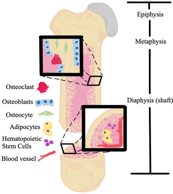
Over the past century, the study of biological processes in the human body has progressed from tissue culture on glass plates to complex 3D models of tissues, organs, and body systems. These dynamic 3D systems have allowed for more accurate recapitulation of human physiology and pathology, which has yielded a platform for disease study with a greater capacity to understand pathophysiology and to assess pharmaceutical treatments. Specifically, by increasing the accuracy with which the microenvironments of disease processes are modeled, the clinical manifestation of disease has been more accurately reproduced in vitro. The application of these models is crucial in all realms of medicine, but they find particular utility in diseases related to the complex bone marrow niche. Osteoblast, osteoclasts, bone marrow adipocytes, mesenchymal stem cells, and red and white blood cells represent some of cells that call the bone marrow microenvironment home. During states of malignant marrow disease, neoplastic cells migrate to and join this niche. These cancer cells both exploit and alter the niche to their benefit and to the patient's detriment. Malignant disease of the bone marrow, both primary and secondary, is a significant cause of morbidity and mortality today. Innovative study methods are necessary to improve patient outcomes. In this review, we discuss the evolution of 3D models and compare them to the preceding 2D models. With a specific focus on malignant bone marrow disease, we examine 3D models currently in use, their observed efficacy, and their potential in developing improved treatments and eventual cures. Finally, we comment on the aspects of 3D models that must be critically examined as systems continue to be optimized so that they can exert greater clinical impact in the future. © 2019 The Authors. JBMR Plus published by Wiley Periodicals, Inc. on behalf of American Society for Bone and Mineral Research.
Keywords: 3D MODELS; BONE MARROW; BONE MARROW ADIPOSE; IN VITRO MODELS; TISSUE ENGINEERING.
© 2019 The Authors. JBMR Plus published by Wiley Periodicals, Inc. on behalf of American Society for Bone and Mineral Research.
Figures
References
-
- Faculty of Biological Sciences, University of Leeds . The histology guide. Cartilage, bone & ossification [Internet]. [Cited 16 March 2019]. Available from: https://www.histology.leeds.ac.uk/bone/bone.php.
-
- Morgan EF, Barnes GL, Einhorn TA. The bone organ system: form and function In Marcus R, Feldman D, Nelson DA, Rosen CJ, eds. Osteoporosis. San Diego, CA: Academic Press; 2008. pp 3–25.Editors.
-
- Cooper DML, Kawalilak CE, Harrison K, Johnston BD, Johnston JD. Cortical bone porosity: what is it, why is it important, and how can we detect it? Curr Osteoporos Rep. 2016;14(5):187–98. - PubMed
-
- Wopenka B, Pasteris JD. A mineralogical perspective on the apatite in bone. Mater Sci Eng C. 2005;25(2):131–43.
Publication types
Grants and funding
LinkOut - more resources
Full Text Sources
Miscellaneous