

Minimal Duration of Tick Attachment Sufficient for Transmission of Infectious Rickettsia rickettsii (Rickettsiales: Rickettsiaceae) by Its Primary Vector Dermacentor variabilis (Acari: Ixodidae): Duration of Rickettsial Reactivation in the Vector Revisited
- PMID: 31687749
- PMCID: PMC7171694
- DOI: 10.1093/jme/tjz191
Minimal Duration of Tick Attachment Sufficient for Transmission of Infectious Rickettsia rickettsii (Rickettsiales: Rickettsiaceae) by Its Primary Vector Dermacentor variabilis (Acari: Ixodidae): Duration of Rickettsial Reactivation in the Vector Revisited
Abstract
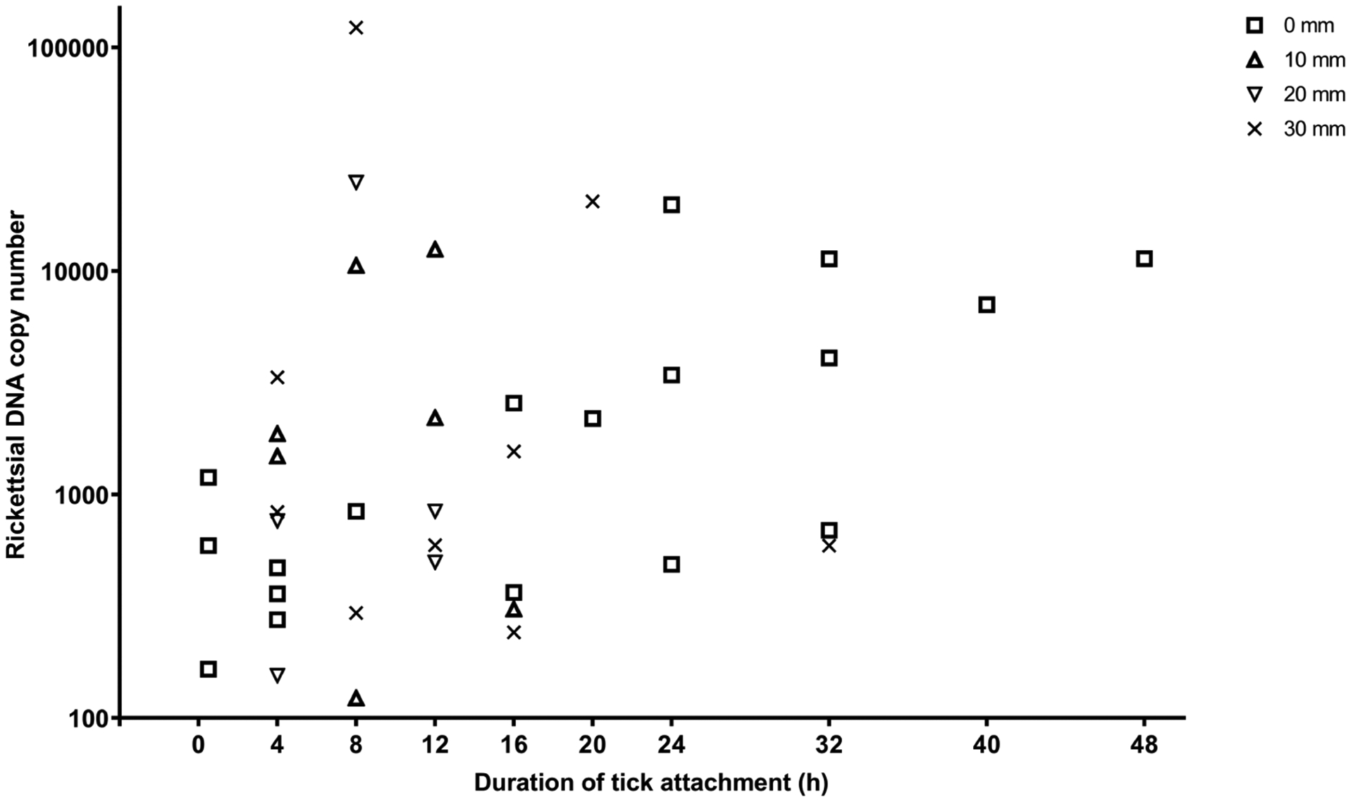
It has been reported that starving ticks do not transmit spotted fever group Rickettsia immediately upon attachment because pathogenic bacteria exist in a dormant, uninfectious state and require time for 'reactivation' before transmission to a susceptible host. To clarify the length of reactivation period, we exposed guinea pigs to bites of Rickettsia rickettsii-infected Dermacentor variabilis (Say) and allowed ticks to remain attached for predetermined time periods from 0 to 48 h. Following removal of attached ticks, salivary glands were immediately tested by PCR, while guinea pigs were observed for 10-12 d post-exposure. Guinea pigs in a control group were subcutaneously inoculated with salivary glands from unfed D. variabilis from the same cohort. In a parallel experiment, skin at the location of tick bite was also excised at the time of tick removal to ascertain dissemination of pathogen from the inoculation site. Animals in every exposure group developed clinical and pathological signs of infection. The severity of rickettsial infection in animals increased with the length of tick attachment, but even attachments for less than 8 h resulted in clinically identifiable infection in some guinea pigs. Guinea pigs inoculated with salivary glands from unfed ticks also became severely ill. Results of our study indicate that R. rickettsii residing in salivary glands of unfed questing ticks does not necessarily require a period of reactivation to precede the salivary transmission and ticks can transmit infectious Rickettsia virtually as soon as they attach to the host.
Keywords: Rickettsia rickettsii; grace period; reactivation; transmission dynamics.
Published by Oxford University Press on behalf of Entomological Society of America 2019.
Figures


 ; 1–
; 1– ; 2–
; 2– ; 3–
; 3– ; 4–
; 4– ; 5–
; 5– ; 6–
; 6– ; 7–
; 7– . **Detection of rickettsial DNA in ear-skin biopsies and/or internal tissues signifies dissemination of R. rickettsii from the site of tick bite or needle-inoculation.
References
-
- Alekseev AN, Burenkova LA, Podboronov VM, and Chunikhin SP. 1995. Bacteriocidal qualities of ixodid tick (Acarina: Ixodidae) salivary cement plugs and their changes under the influence of a viral tick-borne pathogen. J. Med. Entomol 32: 578–582. - PubMed
-
- Alekseev AN, Burenkova LA, Vasilieva IS, Dubinina EV, and Chunikhin SP. 1996. Preliminary studies on virus and spirochete accumulation in the cement plug of ixodid ticks. Exp. Appl. Acarol 20: 713–723. - PubMed
-
- Biggs HM, Barton Behravesh C, Bradley KK, Dahlgren FS, Drexler NA, Dumler JS, Folk SM, Kato CY, Lash RR, Levin ML, et al. 2016. Diagnosis and management of tickborne rickettsial diseases: Rocky Mountain spotted fever and other spotted fever group rickettsioses, ehrlichioses, and anaplasmosis—United States: a practical guide for health-care and public health professionals. MMWR Recomm. Rep 65: 1–48. - PubMed
-
- Binnington KC 1978. Sequential changes in salivary gland structure during attachment and feeding of the cattle tick, Boophilus microplus. Int. J. Parasitol 8: 97–115. - PubMed
Publication types
MeSH terms
Grants and funding
LinkOut - more resources
Full Text Sources
