

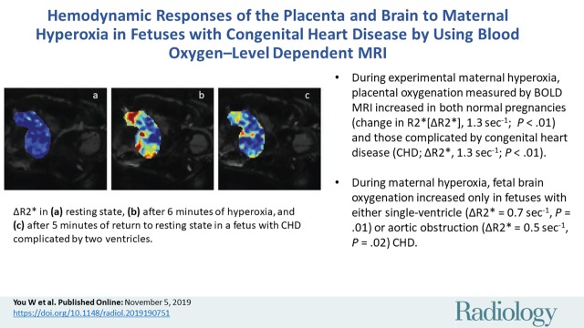
Hemodynamic Responses of the Placenta and Brain to Maternal Hyperoxia in Fetuses with Congenital Heart Disease by Using Blood Oxygen-Level Dependent MRI
- PMID: 31687920
- PMCID: PMC6939742
- DOI: 10.1148/radiol.2019190751
Hemodynamic Responses of the Placenta and Brain to Maternal Hyperoxia in Fetuses with Congenital Heart Disease by Using Blood Oxygen-Level Dependent MRI
Abstract
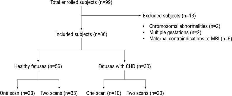
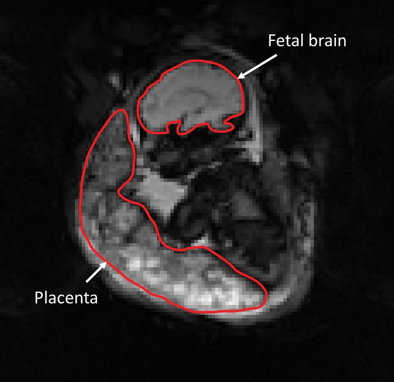
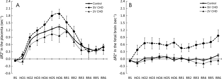
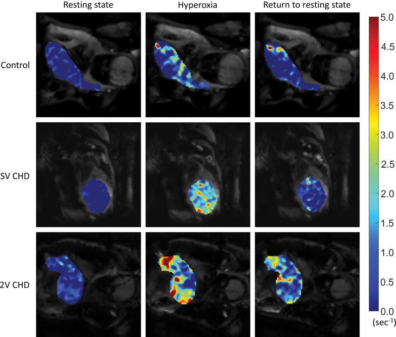
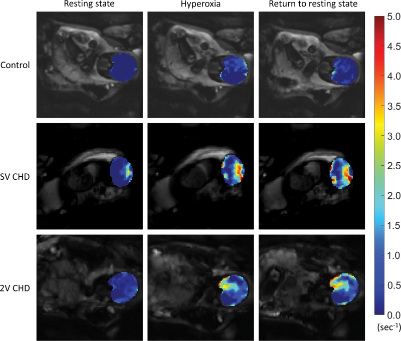
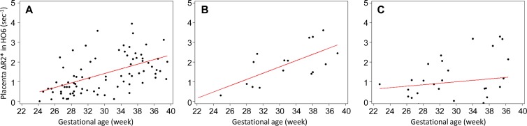
Background Impaired brain development in fetuses with congenital heart disease (CHD) may result from inadequate cerebral oxygen supply in utero. Purpose To test whether fetal cerebral oxygenation can be increased by maternal oxygen administration, effects of maternal hyperoxia on blood oxygenation of the placenta and fetal brain were examined by using blood oxygenation level-dependent (BOLD) functional MRI. Materials and Methods In this prospective study, BOLD MRI was performed in 86 fetuses (56 healthy fetuses and 30 fetuses diagnosed with CHD) between 22 and 39 weeks gestational age (GA) from May 2015 to December 2017, with the following study design: phase I, 2-minute resting state at baseline (room air); phase II, 6-minute maternal hyperoxia with 100% oxygen; and phase III, 5.6-minute return to resting state. After motion correction, the signals were averaged over the placenta and fetal brain and converted to the change in R2* (ΔR2*). Fetuses with CHD were categorized into those with a single ventricle (SV) or two ventricles (TVs) and those with aortic obstruction (AO) or non-AO. Data were analyzed by using generalized linear mixed models controlling for GA and sex. Results Placental ΔR2* increased during maternal hyperoxia in healthy fetuses and fetuses with CHD, but it was higher in SV CHD (mean ΔR2*, 1.3 sec-1 ± 0.1 [standard error; P < .01], 1.9 sec-1 ± 0.2 [P < .01], and 1.0 sec-1 ± 0.3 [P < .01], respectively, for control fetuses, fetuses with SV CHD, and fetuses with TV CHD). Placental ΔR2* during maternal hyperoxia changed with GA in healthy control fetuses and fetuses with SV or AO CHD (ΔR2* per week, 0.1 sec-1 ± 0 [P < .01], 0.2 sec-1 ± 0 [P = .01], and 0.2 sec-1 ± 0 [P = .01], respectively), but not in fetuses with CHD and TV or non-AO. Fetal brain ΔR2* was constant across all phases in healthy control fetuses and fetuses with TV CHD but increased during maternal hyperoxia in fetuses with SV or AO CHD (mean ΔR2*, 0.7 sec-1 ± 0.2 [P = .01] and 0.5 sec-1 ± 0.2 [P = .02], respectively). Conclusion Six minutes of maternal hyperoxia increased placental oxygenation in healthy fetuses and fetuses with congenital heart disease, and it selectively increased cerebral blood oxygenation in fetuses with single ventricle or aortic obstruction. © RSNA, 2019 Online supplemental material is available for this article.
Figures
References
-
- Miller SP, McQuillen PS, Hamrick S, et al. . Abnormal brain development in newborns with congenital heart disease. N Engl J Med 2007;357(19):1928–1938. - PubMed
-
- Donofrio MT, Bremer YA, Schieken RM, et al. . Autoregulation of cerebral blood flow in fetuses with congenital heart disease: the brain sparing effect. Pediatr Cardiol 2003;24(5):436–443. - PubMed
-
- Co-Vu J, Lopez-Colon D, Vyas HV, Weiner N, DeGroff C. Maternal hyperoxygenation: A potential therapy for congenital heart disease in the fetuses? A systematic review of the current literature. Echocardiography 2017;34(12):1822–1833. - PubMed
