. 2019 Nov;27(11):1846-1855.
doi: 10.1002/oby.22607.

Resting-State Brain Connectivity Predicts Weight Loss and Cognitive Control of Eating Behavior After Vertical Sleeve Gastrectomy

Affiliations

Resting-State Brain Connectivity Predicts Weight Loss and Cognitive Control of Eating Behavior After Vertical Sleeve Gastrectomy

Hilâl Cerit et al. Obesity (Silver Spring). 2019 Nov.

Abstract

Objective: The effects of sleeve gastrectomy (SG) on functional connectivity (FC) and associations with weight loss and eating-related cognitive control were investigated.

Methods: In a longitudinal study, 14 SG patients (13 female; 42.1 presurgery BMI) completed study visits 1 month pre surgery and 12 months post surgery. Patients completed the Dutch Eating Behavior Questionnaire and resting-state functional magnetic resonance imaging scanning to measure FC. Data were analyzed using a seed-to-voxel approach in the CONN Toolbox to investigate pre-/postsurgery changes (n = 12) and to conduct predictive analysis (n = 14).

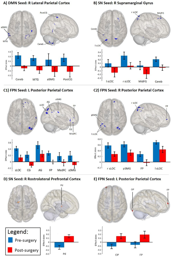
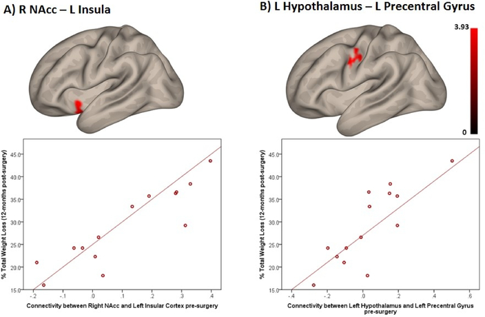
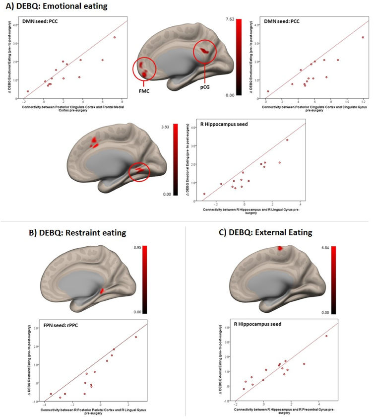
Results: Seed-to-voxel analysis revealed changes in magnitude (decreases) and directionality (positively correlated to anticorrelated) of FC pre to post surgery within and between default mode network, salience network, and frontoparietal network nodes [Family-Wise Error (FWE) corrected at P < 0.05]. Baseline FC of the nucleus accumbens (with insula) and hypothalamus (with precentral gyrus) predicted 12-month post-SG % total weight loss (FWE-P < 0.05). Baseline FC of the hippocampus, frontoparietal network, and default mode network nodes predicted improvement in cognitive control of eating behavior 12 months after SG (FWE-P < 0.05).

Conclusions: Our findings demonstrate changes in FC magnitude and directionality post versus pre surgery within and between resting-state networks and frontal, paralimbic, and visual areas in SG patients. Baseline FC predicted weight loss and changes in cognitive control of food intake behavior at 12 months. These could serve as predictive biomarkers for bariatric surgery.


Conflict of interest statement

Disclosure

The authors have no conflicts of interest to disclose. The findings and conclusions in this report are those of the authors and do not necessarily represent the views of the National Institutes of Health.

Figures

Figure 1.
Figure 1.
Quantitative connectivity of DMN, SN and FPN with significant clusters (derived from seed-to-voxel whole-brain correlation analyses; whole brain height threshold of p<0.001, cluster-size p-FDR-corrected threshold of p<0.05). Bar graphs indicate average connectivity values (i.e., mean connectivity z-scores) extracted from largest clusters for each condition (i.e., pre- and post-surgery) to assess the directionality and magnitude of the observed changes. (A) Change in directionality pre- (positively correlated) to post-surgery (anticorrelated) between Default Mode Network (Seed: Right Lateral Parietal Cortex) and Right Cerebellum (Cereb), Left Middle Temporal Gyrus (MTG), Left Supramarginal Gyrus, anterior division (aSMG), and Right Postcentral Gyrus (PostCG). (B) Change in directionality pre- (positively correlated) to post-surgery (anticorrelated) between Salience Network (Seed: Right Supramarginal Gyrus) and Left Lateral Occipital Cortex, superior division (l sLOC), Right Middle Frontal Gyrus (MidFG) and Left Cerebellum (Cereb). This same SN seed demonstrated reduced magnitude of positive correlation post-surgery (vs. pre-surgery) with the Right Lateral Occipital Cortex, superior division (r sLOC). (C1) Reduced magnitude of positive correlations post-surgery (vs. pre-surgery) between Frontoparietal Network (Seed: Left Posterior Parietal Cortex) and Right Lateral Occipital Cortex, superior division (sLOC) and Right Angular Gyrus (AG). This same FPN seed showed a change in directionality pre- (positively correlated) to post-surgery (anticorrelated) with the Cingulate Gyrus, anterior division (CG), Right Frontal Pole (FP), Frontal Medial Cortex (MedFC), and Right Supramarginal Gyrus, anterior division (aSMG). (C2) Reduced magnitude of positive correlations post-surgery (vs. pre-surgery) between Frontoparietal Network (Seed: Right Posterior Parietal Cortex) and Lateral Occipital Cortex, superior division (sLOC; Left and Right), Left Supramarginal Gyrus, posterior division (pSMG). This same FPN seed demonstrated anticorrelations post-surgery (vs. positive correlations pre-surgery) with the Right Frontal Pole (FP). (D) Change in directionality pre- (anticorrelated) to post-surgery (positively correlated) between Salience Network (Seed: Right Rostrolateral Prefrontal Cortex) and Pallidum (Pd) (E) Change in directionality pre- (anticorrelated) to post-surgery (positively correlated) between Frontoparietal Network (Seed: Left Posterior Parietal Cortex) and Left Occipital pole (OP), Left Frontal Pole (FP).
Figure 2.
Figure 2.
Positive association between pre-surgery functional connectivity and % Total Weight Loss 12-months post-surgery (note: color scale indicates T-values; the X-axis indicate average connectivity values). (A) Right Nucleus Accumbens and Left Insular Cortex (p-FDR-corrected<0.001). (B) Left Hypothalamus and Left Precentral Gyrus (p-FDR-corrected=0.024).
Figure 3.
Figure 3.
Positive association between pre-surgery functional connectivity (in the DMN, FPN and hippocampus) and increase in cognitive control of eating from pre- to post surgery (note: color scale indicates T-values; X-axis on scatterplot indicates average connectivity values). (A) DEBQ Emotional Eating subscale associated with FC between the Posterior Cingulate Cortex (DMN) with Frontal Medial Cortex (FMC; L/R) (p-FDR corrected<0.001) and PCC (DMN) with Cingulate Gyrus, posterior division (pCG) (p-FDR-corrected<0.001) (upper panel) and with FC between the right Hippocampus with R Lingual Gyrus/Paracingulate Gyrus (p-FDR-corrected=0.019) (lower panel). (B) DEBQ Restraint Eating subscale associated with FC between the Right Posterior Parietal Cortex (FPN) with Right Lingual Gyrus (p-FDR-corrected=0.037). (C) DEBQ External Eating subscale associated with FC between the Right Hippocampus with Right Precentral Gyrus (p-FDR-corrected=0.018).

References

    1. Chang S, Stoll C, Song J, Varela J, Eagon C, Colditz G. The effectiveness and risks of bariatric surgery: An updated systematic review and meta-analysis, 2003–2012. JAMA Surg 2014;149: 275–287. - PMC - PubMed
    1. Courcoulas AP, Christian NJ, Belle SH, et al. Weight change and health outcomes at 3 years after bariatric surgery among individuals with severe obesity. JAMA 2013;310: 2416–2425. - PMC - PubMed
    1. Holsen L, Davidson P, Cerit H, Hye T, Moondra P, Haimovici F et al. Neural predictors of 12-month weight loss outcomes following bariatric surgery. Int J Obes 2018;42: 785–793. - PMC - PubMed
    1. Ness A, Bruce J, Bruce A, Aupperle R, Lepping R, Martin L, et al. Pre-surgical cortical activation to food pictures is associated with weight loss following bariatric surgery. Surg Obes Relat Dis 2014;10: 1188–1195. - PubMed
    1. Ochner CN, Stice E, Hutchins E, Afifi L, Geliebter A, Hirsch J, et al. Relation between changes in neural responsivity and reductions in desire to eat high-calorie foods following gastric bypass surgery. Neuroscience 2012;209: 128–135. - PMC - PubMed

Publication types

MeSH terms