. 2019 Dec 1;317(6):E1182-E1192.
doi: 10.1152/ajpendo.00338.2019. Epub 2019 Nov 5.

The gut microbiota is a major regulator of androgen metabolism in intestinal contents

Affiliations

The gut microbiota is a major regulator of androgen metabolism in intestinal contents

Hannah Colldén et al. Am J Physiol Endocrinol Metab. .

Abstract

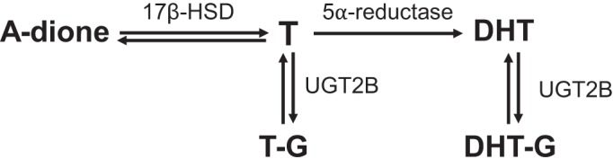
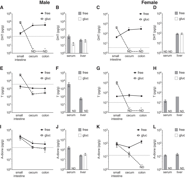
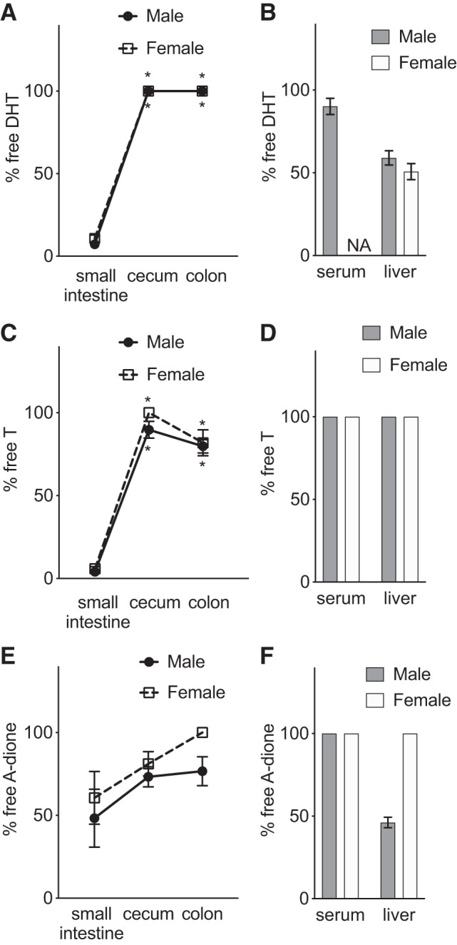
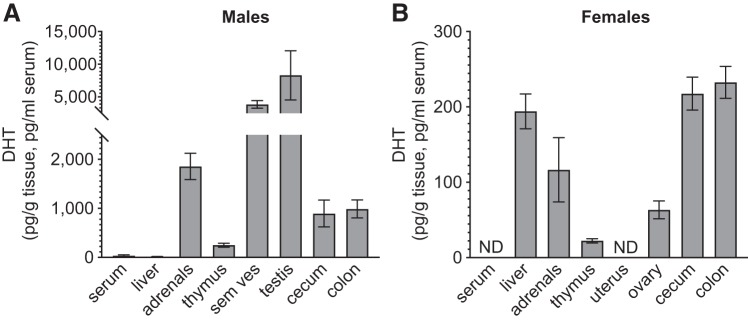
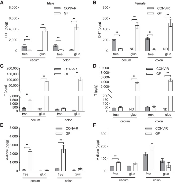
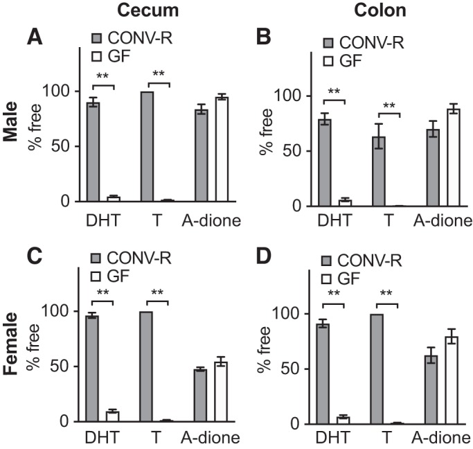
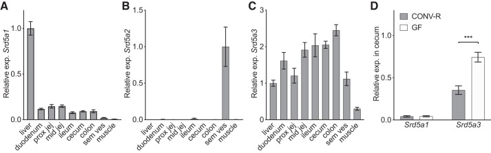
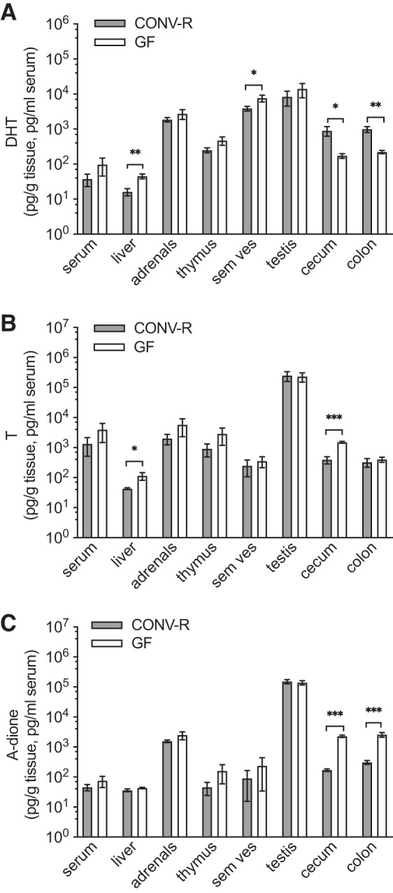
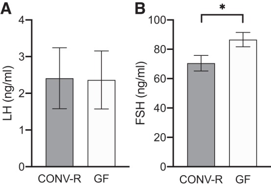
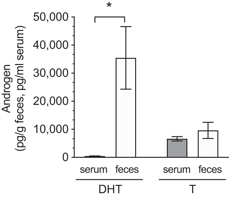
Androgens exert important effects both in androgen-responsive tissues and in the intestinal tract. To determine the impact of the gut microbiota (GM) on intestinal androgen metabolism, we measured unconjugated (free) and glucuronidated androgen levels in intestinal contents from the small intestine, with a low bacterial density, and from cecum and colon, with a high bacterial density. Using a specific, sensitive gas chromatography-tandem mass spectrometry method, we detected high levels of glucuronidated testosterone (T) and dihydrotestosterone (DHT) in small intestinal content of mice of both sexes, whereas in the distal intestine we observed remarkably high levels of free DHT, exceeding serum levels by >20-fold. Similarly, in young adult men high levels of unconjugated DHT, >70-fold higher than in serum, were detected in feces. In contrast to mice with a normal GM composition, germ-free mice had high levels of glucuronidated T and DHT, but very low free DHT levels, in the distal intestine. These findings demonstrate that the GM is involved in intestinal metabolism and deglucuronidation of DHT and T, resulting in extremely high free levels of the most potent androgen, DHT, in the colonic content of young and healthy mice and men.

Keywords: dihydrotestosterone; glucuronidation; gut bacteria; intracrinology; testosterone.


Conflict of interest statement

No conflicts of interest, financial or otherwise, are declared by the authors.

Figures

Fig. 1.
Fig. 1.
Schematic representation of androgen metabolism. The androgen precursor androstenedione (A-dione) can be reversibly metabolized to testosterone (T) by 17β-hydroxysteroid dehydrogenase enzymes (17β-HSD). T can then be reduced by 5α-reductase enzymes to the more potent androgen dihydrotestosterone (DHT). Both T and DHT can be glucuronidated (-G) in the liver by uridine diphosphate-glucuronosyltransferase (UGT) 2B enzymes to make them more water soluble and facilitate excretion. Glucuronidation is reversible.
Fig. 2.
Fig. 2.
Comparison of glucuronidated and unconjugated androgens in contents of small and distal intestine of mice with normal gut microbiota. Unconjugated (free) and glucuronidated (gluc) androgen levels were measured by gas chromatography-tandem mass spectrometry in different parts of the intestine, serum, and liver of 8-wk-old C57BL/6 mice. A–D: dihydrotestosterone (DHT) in males (A and B) and females (C and D). E–H: testosterone (T) in males (E and F) and females (G and H). I–L: the androgen precursor androstenedione (A-dione) in males (I and J) and females (K and L). Values are shown as means ± SE; n = 7 mice. ND, not detectable; dotted horizontal line denotes the corresponding serum free level. In serum the unit of measurement is picograms per milliliter. To compare cecum and colon with small intestine, Wilcoxon matched-pairs signed-rank test was employed. *P < 0.05 vs. small intestine.
Fig. 3.
Fig. 3.
Comparison of unconjugated androgen fraction in small and distal intestine of mice with normal gut microbiota: unconjugated fraction (% free) of dihydrotestosterone (DHT; A and B), testosterone (T; C and D), and androstenedione (A-dione; E and F) in parts of the intestine, serum, and liver of 8-wk-old male and female C57BL/6 mice. Values are shown as means ± SE; n = 7 mice. NA, not applicable. To compare cecum and colon with small intestine, Wilcoxon matched-pairs signed-rank test was employed. *P < 0.05 vs. small intestine.
Fig. 4.
Fig. 4.
Comparison of unconjugated dihydrotestosterone (DHT) levels in intestine and extraintestinal tissues and serum of mice with normal gut microbiota: DHT levels in intestinal contents, serum, and tissues of 8-wk-old male (A) and female (B) C57BL/6 mice. Values below lower limit of quantification (LLOQ) are set to LLOQ; if no detectable levels in group, the level is denoted not detectable (ND). Values are shown as means ± SE; n = 10 per group. Sem ves, seminal vesicles.
Fig. 5.
Fig. 5.
Comparison of glucuronidated and unconjugated androgens in intestinal contents of conventionally raised (CONV-R) and germ-free (GF) mice: unconjugated (free) and glucuronidated (gluc) dihydrotestosterone (DHT; A and B), testosterone (T; C and D), and androstenedione (A-dione; E and F) levels in intestinal contents of 8-wk-old male (A, C, E) and female (B, D, F) CONV-R and GF mice. Values below lower limit of quantification (LLOQ) are set to LLOQ or denoted not detectable (ND). Values are shown as means ± SE; n = 10 per group. To compare the 2 groups, the Mann–Whitney U test was employed. **P < 0.01.
Fig. 6.
Fig. 6.
Comparison of unconjugated androgen fractions in the intestine of conventionally raised (CONV-R) and germ-free (GF) mice: unconjugated fraction (% free) of dihydrotestosterone (DHT), testosterone (T) and androstenedione (A-dione) in cecal (A and C) and colonic (B and D) contents of 8-wk-old male (A and B) and female (C and D) CONV-R and GF C57BL/6 mice. Values are shown as means ± SE; n = 10 per group. To compare the 2 groups, the Mann–Whitney U test was employed. **P < 0.01.
Fig. 7.
Fig. 7.
Gene expression of 5α-reductases (Srd5a1, Srd5a2, Srd5a3) in the intestinal tract. A–C: gene expression of 5α-reductase type 1 (A), type 2 (B), and type 3 (C) in tissues of 8-wk-old male C57BL/6 mice (n = 7). D: gene expression of Srd5a1 and Srd5a3 in cecum of 8-wk-old germ-free (GF) and conventionally raised (CONV-R) male mice (n = 10 per group). Relative expression was calculated with 18S as reference gene. Values are shown as means ± SE. Significant differences between GF and CONV-R according to Student’s 2-tailed unpaired t test, ***P < 0.0001. Prox jej, proximal jejunum; mid jej, mid jejunum; sem ves, seminal vesicles.
Fig. 8.
Fig. 8.
Comparison of unconjugated (free) androgen levels in intestine and extraintestinal tissues of conventionally raised (CONV-R) and germ-free (GF) male mice: dihydrotestosterone (DHT; A), testosterone (T; B) and androstenedione (A-dione; C). Values below lower limit of quantification (LLOQ) are set to LLOQ. Values are shown as means ± SE; n = 10 per group. Significance: *P < 0.05; **P < 0.01; ***P < 0.0001 according to Student’s 2-tailed unpaired t test. Sem ves, seminal vesicles.
Fig. 9.
Fig. 9.
Serum levels of gonadotropins in conventionally raised (CONV-R) and germ-free (GF) male mice: luteinizing hormone (LH; A) and follicle-stimulating hormone (FSH; B). Values are shown as means ± SE; n = 8 per group. Significance: *P < 0.05 according to Student’s 2-tailed unpaired t test.
Fig. 10.
Fig. 10.
Comparison of unconjugated androgen levels in feces and serum of young adult men. Unconjugated (free) dihydrotestosterone (DHT) and testosterone (T) levels were measured by gas chromatography-tandem mass spectrometry. Values are shown as means ± SE; n = 8 subjects. *P = 0.016 according to Student’s 2-tailed paired t test.

References

    1. Abotchie PN, Vernon SW, Du XL. Gender differences in colorectal cancer incidence in the United States, 1975-2006. J Womens Health (Larchmt) 21: 393–400, 2012. doi:10.1089/jwh.2011.2992. - DOI - PMC - PubMed
    1. Al-Asmakh M, Stukenborg JB, Reda A, Anuar F, Strand ML, Hedin L, Pettersson S, Söder O. The gut microbiota and developmental programming of the testis in mice. PLoS One 9: e103809, 2014. doi:10.1371/journal.pone.0103809. - DOI - PMC - PubMed
    1. Amos-Landgraf JM, Heijmans J, Wielenga MC, Dunkin E, Krentz KJ, Clipson L, Ederveen AG, Groothuis PG, Mosselman S, Muncan V, Hommes DW, Shedlovsky A, Dove WF, van den Brink GR. Sex disparity in colonic adenomagenesis involves promotion by male hormones, not protection by female hormones. Proc Natl Acad Sci USA 111: 16514–16519, 2014. doi:10.1073/pnas.1323064111. - DOI - PMC - PubMed
    1. Anderson RC, Newton CL, Anderson RA, Millar RP. Gonadotropins and their analogs: current and potential clinical applications. Endocr Rev 39: 911–937, 2018. doi:10.1210/er.2018-00052. - DOI - PubMed
    1. Ba ZF, Yokoyama Y, Toth B, Rue LW 3rd, Bland KI, Chaudry IH. Gender differences in small intestinal endothelial function: inhibitory role of androgens. Am J Physiol Gastrointest Liver Physiol 286: G452–G457, 2004. doi:10.1152/ajpgi.00357.2003. - DOI - PubMed

Publication types

MeSH terms

LinkOut - more resources