Clinical Trial
. 2020 Feb 3;130(2):733-747.
doi: 10.1172/JCI121127.

Off-the-shelf EBV-specific T cell immunotherapy for rituximab-refractory EBV-associated lymphoma following transplantation

Affiliations
Clinical Trial

Off-the-shelf EBV-specific T cell immunotherapy for rituximab-refractory EBV-associated lymphoma following transplantation

Susan Prockop et al. J Clin Invest. .

Abstract

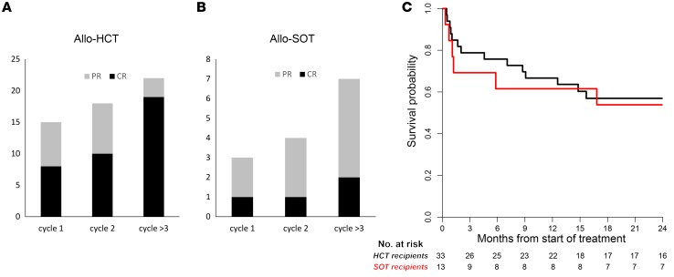
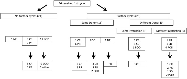
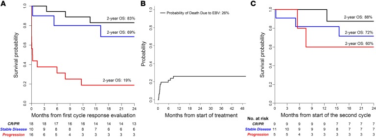
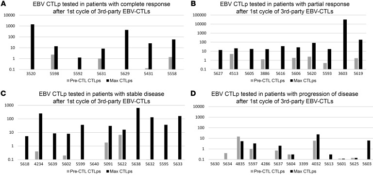
BACKGROUNDAdoptive transfer of donor-derived EBV-specific cytotoxic T-lymphocytes (EBV-CTLs) can eradicate EBV-associated lymphomas (EBV-PTLD) after transplantation of hematopoietic cell (HCT) or solid organ (SOT) but is unavailable for most patients.METHODSWe developed a third-party, allogeneic, off-the-shelf bank of 330 GMP-grade EBV-CTL lines from specifically consented healthy HCT donors. We treated 46 recipients of HCT (n = 33) or SOT (n = 13) with established EBV-PTLD, who had failed rituximab therapy, with third-party EBV-CTLs. Treatment cycles consisted of 3 weekly infusions of EBV-CTLs and 3 weeks of observation.RESULTSEBV-CTLs did not induce significant toxicities. One patient developed grade I skin graft-versus-host disease. Complete remission (CR) or sustained partial remission (PR) was achieved in 68% of HCT recipients and 54% of SOT recipients. For patients who achieved CR/PR or stable disease after cycle 1, one year overall survival was 88.9% and 81.8%, respectively. In addition, 3 of 5 recipients with POD after a first cycle who received EBV-CTLs from a different donor achieved CR or durable PR (60%) and survived longer than 1 year. Maximal responses were achieved after a median of 2 cycles.CONCLUSIONThird-party EBV-CTLs of defined HLA restriction provide safe, immediately accessible treatment for EBV-PTLD. Secondary treatment with EBV-CTLs restricted by a different HLA allele (switch therapy) can also induce remissions if initial EBV-CTLs are ineffective. These results suggest a promising potential therapy for patients with rituximab-refractory EBV-associated lymphoma after transplantation.TRIAL REGISTRATIONPhase II protocols (NCT01498484 and NCT00002663) were approved by the Institutional Review Board at Memorial Sloan Kettering Cancer Center, the FDA, and the National Marrow Donor Program.FUNDINGThis work was supported by NIH grants CA23766 and R21CA162002, the Aubrey Fund, the Claire Tow Foundation, the Major Family Foundation, the Max Cure Foundation, the Richard "Rick" J. Eisemann Pediatric Research Fund, the Banbury Foundation, the Edith Robertson Foundation, and the Larry Smead Foundation. Atara Biotherapeutics licensed the bank of third-party EBV-CTLs from Memorial Sloan Kettering Cancer Center in June 2015.

Keywords: Cancer immunotherapy; Immunology; T cells; Transplantation.


Conflict of interest statement

Conflict of interest: ED and ROR had consultancy agreements with Atara Biotherapeutics. ED and ROR are inventors on technology referenced in this work (SK2013-71 [patient ID], Proprietary Cell Banks for Use in 3rd-Party EBV-Specific T Cell Therapy; and SK2018-122 [patient ID], Methods of Selecting T Cell Lines for Adoptive Cellular Therapy). SP is an inventor on technology referenced in this work (SK2013-71 and SK2018-122) and has waived rights to revenue generated from these inventions. Memorial Sloan Kettering Cancer Center (MSK), which owns the technology, has licensed this technology to Atara, and MSK has interests in Atara through this licensing arrangement.

Figures

Figure 1
Figure 1. Characterization of 55 EBV-specific cytotoxic T cells infused.
All T cell lines, including those predominantly containing CD4+ T cells, demonstrated EBV-specific cytotoxic activity against autologous EBV-BLCLs and did not kill NK cell–sensitive targets (K562), EBV-negative autologous or recipient-derived PHA blasts, or HLA-mismatched EBV-BLCLs. (A) Phenotype (CD3, CD8, CD4, and NK). (B) Cytotoxic activity of EBV-specific T cell lines against autologous BLCLs (circles), autologous PHA blasts (squares), mismatched targets (triangles), and NK-sensitive K562 targets (inverted triangles). (C) EBV-CTLp frequency and alloreactive CTL precursor (allo-CTLp) frequency in lines infused to treat patients. In limiting dilution assays, the EBV-CTLs contained a median of 6323.5 EBV-CTLps per 106 cells (range, 2.5–76,982 EBV-CTLps per 106 cells), and, in response to irradiated fully allogeneic PBMCs, generated low or undetectable alloreactive CTLps (median 1.2, range 0–27.4 allo-CTLps per 106 cells). Error bars indicate ± SD.
Figure 2
Figure 2. Number of cycles to best response (CR or PR), and survival of patients with HCT or SOT.
(A) Patients with EBV lymphoma after HCT. (B) Patients with EBV lymphoma after SOT. Patients achieving a CR (black) after the first cycle of EBV-CTLs included 8 of 33 HCT recipients and 1 of 13 SOT recipients. An additional 9 patients (7 HCT, 2 SOT) achieved a PR (gray), and 10 had SD. Thus, the overall response (CR + PR) after cycle 1 was 39% (18/46). With subsequent cycles of EBV-CTLs the response rate increased to 22 of 33 HCT (68%) and 7 of 13 SOT (54%) recipients. (C) Kaplan-Meier probabilities of survival for HCT and SOT patients at 2 years were 57% and 54%, respectively.
Figure 3
Figure 3. Flowchart of treatment and responses for patients treated for EBV-PTLD with third-party EBV-CTLs.
DOD, dead of disease.
Figure 4
Figure 4. Overall survival at 2 years.
(A) Survival of all patients based on evaluation of response to the first cycle of third-party EBV-CTLs. (B) Cumulative probability of death due to EBV lymphoma. (C) Survival of patients who received subsequent cycles of third-party EBV-CTLs, based on status of the EBV lymphoma immediately prior to initiation of the second cycle of EBV-CTLs. OS, overall survival.
Figure 5
Figure 5. EBV-CTLp frequency after first cycle of adoptive therapy with third-party EBV-CTLs.
Expansions could be detected in patients with responses as well as those with stable disease. Individual patients are demonstrated by distinctly colored lines. (A) EBV-CTLps tested in patients with complete response after first cycle of third-party EBV-CTLs. (B) EBV-CTLps tested in patients with partial response after first cycle of third-party EBV-CTLs. (C) EBV-CTLps tested in patients with stable disease after first cycle of third-party EBV-CTLs. (D) EBV-CTLps tested in patients with POD after first cycle of third-party EBV-CTLs.
Figure 6
Figure 6. Response to EBV-CTLs restricted by either HLA-A*1101 or HLA-B*4403.
(A) High-resolution typing of the patient and the origin of the EBV+ lymphoma and of the 4 EBV-CTL lines successively infused. Blue type indicates the restricting HLA allele of the EBV-CTL line. (B) Time course of EBV lymphoma and response to successive EBV-CTL lines (EBV PCR as an additional marker of disease). (C) Successive PET scans of disease progression and regression. (D) Distinctive pattern of STRs in EBV-CTLs. Top: Baseline host prior to CTL infusion. Middle: Third-party EBV-CTL donor. Bottom: EBV-specific T cells in blood 32 days after initial infusion and 16 days after third infusion in cycle 1 of EBV-CTLs from donor D, prior to cycle 2. Post-CTL-infusion specimen demonstrating the presence of CTL donor D cells (arrows indicate unique peaks corresponding to donor). s/p, status/post.
Figure 7
Figure 7. Detection of third-party donor EBV-CTLs using short tandem repeat analysis in 2 patients.
(A) HCT recipient. (B) SOT recipient. Baseline host prior to CTL infusion, third-party EBV-CTL donor, and post-CTL-infusion specimens demonstrating the presence of CTL donor cells at serial time points after infusion (arrows indicate unique peaks corresponding to donor). In the SOT patient, third-party donor EBV-CTLs were still detectable 23.7 months after the last infusion.

Comment in

References

    1. Singavi AK, Harrington AM, Fenske TS. Post-transplant lymphoproliferative disorders. Cancer Treat Res. 2015;165:305–327. - PubMed
    1. Hoegh-Petersen M, et al. High incidence of post transplant lymphoproliferative disorder after antithymocyte globulin-based conditioning and ineffective prediction by day 28 EBV-specific T lymphocyte counts. Bone Marrow Transplant. 2011;46(8):1104–1112. doi: 10.1038/bmt.2010.272. - DOI - PubMed
    1. San-Juan R, et al. Epstein-Barr virus-related post-transplant lymphoproliferative disorder in solid organ transplant recipients. Clin Microbiol Infect. 2014;20(suppl 7):109–118. - PubMed
    1. Morscio J, Dierickx D, Tousseyn T. Molecular pathogenesis of B-cell posttransplant lymphoproliferative disorder: what do we know so far? Clin Dev Immunol. 2013;2013:150835. - PMC - PubMed
    1. Petrara MR, Giunco S, Serraino D, Dolcetti R, De Rossi A. Post-transplant lymphoproliferative disorders: from epidemiology to pathogenesis-driven treatment. Cancer Lett. 2015;369(1):37–44. doi: 10.1016/j.canlet.2015.08.007. - DOI - PubMed

Publication types

MeSH terms

Associated data