. 2019 Dec 13;294(50):19081-19098.
doi: 10.1074/jbc.RA119.011153. Epub 2019 Nov 5.

Functional diversification of the chemical landscapes of yeast Sec14-like phosphatidylinositol transfer protein lipid-binding cavities

Affiliations

Functional diversification of the chemical landscapes of yeast Sec14-like phosphatidylinositol transfer protein lipid-binding cavities

Ashutosh Tripathi et al. J Biol Chem. .

Erratum in

Abstract

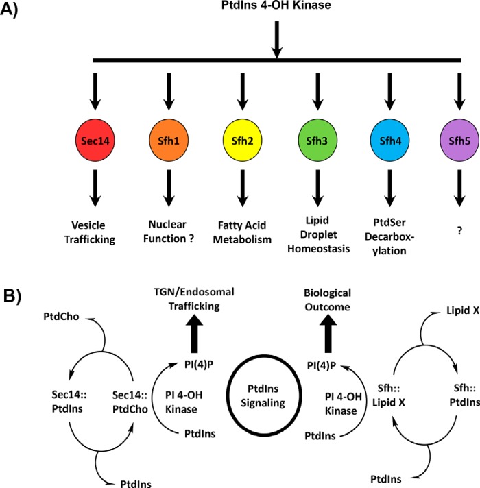
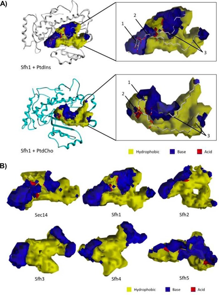
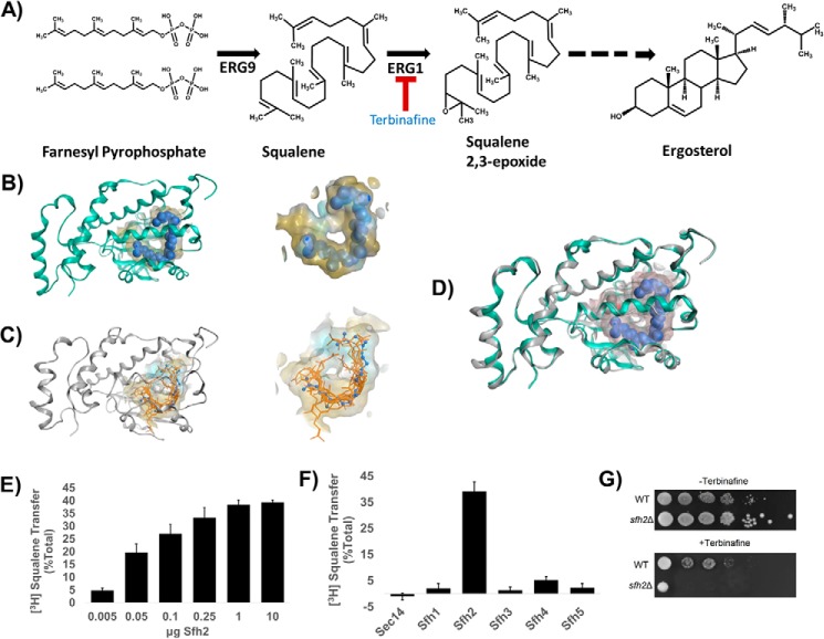
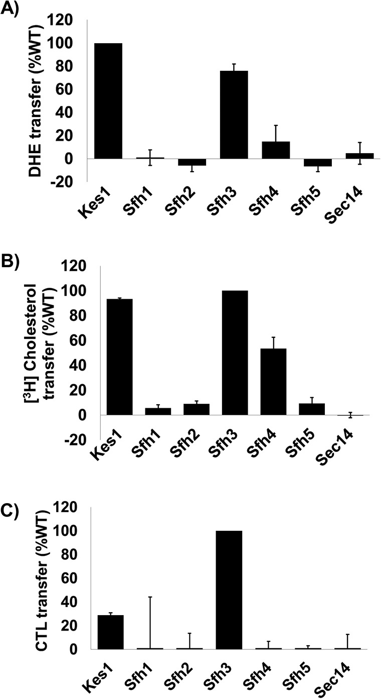
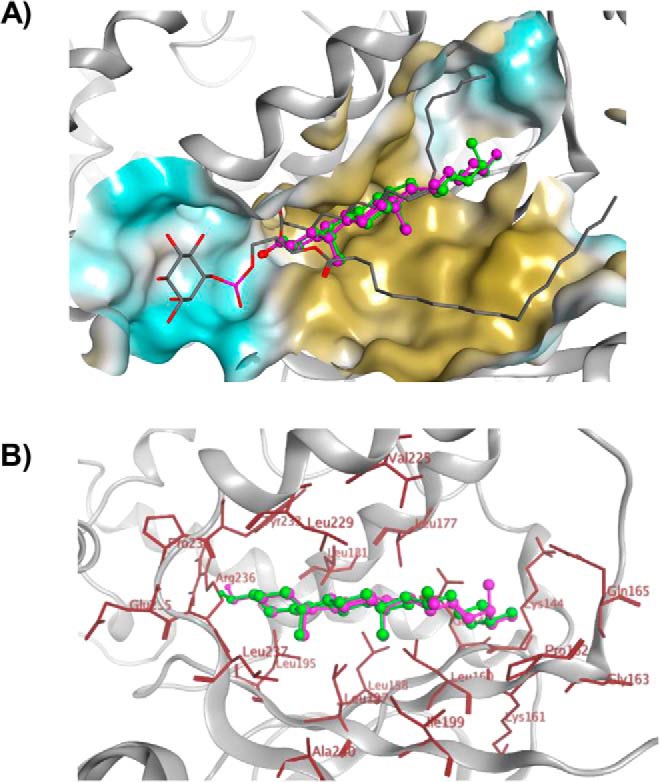
Phosphatidylinositol-transfer proteins (PITPs) are key regulators of lipid signaling in eukaryotic cells. These proteins both potentiate the activities of phosphatidylinositol (PtdIns) 4-OH kinases and help channel production of specific pools of phosphatidylinositol 4-phosphate (PtdIns(4)P) dedicated to specific biological outcomes. In this manner, PITPs represent a major contributor to the mechanisms by which the biological outcomes of phosphoinositide are diversified. The two-ligand priming model proposes that the engine by which Sec14-like PITPs potentiate PtdIns kinase activities is a heterotypic lipid-exchange cycle where PtdIns is a common exchange substrate among the Sec14-like PITP family, but the second exchange ligand varies with the PITP. A major prediction of this model is that second-exchangeable ligand identity will vary from PITP to PITP. To address the heterogeneity in the second exchange ligand for Sec14-like PITPs, we used structural, computational, and biochemical approaches to probe the diversities of the lipid-binding cavity microenvironments of the yeast Sec14-like PITPs. The collective data report that yeast Sec14-like PITP lipid-binding pockets indeed define diverse chemical microenvironments that translate into differential ligand-binding specificities across this protein family.

Keywords: Sec14-domain; cavity mapping; computational biology; lipid metabolism; phosphatidylinositol transfer proteins; phosphoinositide; signaling; squalene; sterol.


Conflict of interest statement

The authors declare that they have no conflicts of interest with the contents of this article

Figures

Figure 1.
Figure 1.
Sec14-like PITPs and diversification of PtdIns(4)P signaling. A illustrates the concept that the biological outcome of PtdIns(4)P signaling in yeast is primarily determined by the Sec14-like PITP that stimulates activity of a PtdIns 4-OH kinase to produce a functionally channeled PtdIns(4)P pool. This strategy is a major mechanism for diversifying PtdIns(4)P signaling. B depicts a heterotypic PtdIns/“second lipid” exchange cycle where the second lipid (lipid X) is a priming lipid that potentiates presentation of PtdIns to the lipid kinase, thereby rendering the PtdIns a superior substrate for the kinase resulting in stimulation of PtdIns(4)P production (3, 4, 6). At left is shown the concept for the Sec14 PtdIns/PtdCho-exchange cycle where PtdIns(4)P is channeled to promotion of membrane trafficking from the TGN/endosomal system. At right is shown the general case proposed for Sec14-like PITPs that fail to bind PtdCho and for whom identification of the exchangeable lipid X is sought.
Figure 2.
Figure 2.
Structural features of Sec14 orthologs. A, α-carbon backbones of the basic folds of open conformers of Sec14 orthologs from the indicated fungal species: S. cerevisiae (blue); C. albicans (orange); Neurospora crassa (pink); C. dubliniensis (magenta); C. glabrata (yellow); K. lactis (cyan); A. nidulans (brown); and S. pombe (teal). At center is a structural overlay of the collective α-carbon backbones onto that of Sec14 that highlights the high overall conservation of the basic fold across large evolutionary distances. B, electrostatic potential surfaces of open conformers Sec14 and Sec14 orthologs from the indicated fungal species are shown. The en face orientation illustrates the open gating helices and the exposed lipid-binding cavities. Surfaces with electropositive potential are rendered blue, and electronegative surfaces are highlighted in red.
Figure 3.
Figure 3.
Structural features of Sec14 paralogs, the Sec14-like Sfh proteins. A, α-carbon backbones of the basic folds of open conformers of Sec14 and the Sec14-like Sfh PITPs from S. cerevisiae: Sec14 (blue); Sfh1 (orange); Sfh2 (pink); Sfh3 (magenta); Sfh4 (cyan); and Sfh5 (teal). At center is a structural overlay of the collective α-carbon backbones superimposed onto that of Sec14 that highlights the overall conservation of the basic Sec14-fold across the yeast Sfh1 family. B, electrostatic potential surfaces of open conformers Sec14 and indicated Sfh PITPs are shown. The en face orientation illustrates the open-gating helices and the exposed lipid-binding cavities and is labeled as “front,” and the opposite side of the molecule is identified as “back.” Surfaces with electropositive potential are rendered blue, and the electronegative surfaces are highlighted in red.
Figure 4.
Figure 4.
Structural barcodes for PtdIns- and PtdCho-binding in Sec14 orthologs and Sec14-like Sfh proteins. A, structural overlay of the PtdIns-binding (left panel) and PtdCho-binding barcode residues (right panel) from Sec14 orthologs superimposed onto those of Sec14. PtdIns-binding barcode residues are shown in blue stick model with PtdIns rendered in green ball and stick. The PtdCho-binding barcode residues are shown in orange stick model, and PtdCho is rendered in green ball and stick. Both barcodes are structurally preserved in Sec14 orthologs across large evolutionary distances. B, structural overlay of the PtdIns-binding (left panel) and PtdCho-binding barcode residues (right panel) from the indicated Sec14-like Sfh PITPs superimposed onto those of Sec14. PtdIns- and PtdCho-binding barcode residues are shown in a stick model using the color code of Fig. 3A to identify residues of individual Sfh proteins. PtdIns is rendered in green ball and stick. The PtdCho-binding barcode regions are shown using the color code of Fig. 3A to identify residues of individual Sfh proteins. PtdCho is rendered in green ball and stick. Although the PtdIns-binding barcodes are structurally well-preserved in the Sfh proteins, the PtdCho barcode is highly divergent (with the exception of Sfh1).
Figure 5.
Figure 5.
VICE/HINT mapping of the Sec14 and Sfh PITP lipid-binding cavities. A, VICE-calculated binding cavity of Sfh1 was mapped with HINT complementary maps, and the PtdIns (top) and PtdCho (bottom) headgroup-binding regions are illustrated. PtdIns is bound in the cavity at the interface between the LBD and tripod motifs. The inositol headgroup (1) and glycerol backbone-binding region (2) are localized in distinctive polar microenvironments of the cavity surface. The acyl chains (3) pack into the largely hydrophobic LBD. PtdCho binds the cavity at the interface between the LBD and tripod motifs. The choline headgroup (1) is located in a hydrophobic area in the near vicinity of an extensive polar region. The PtdCho phosphate moiety (2) is coordinated in a polar surface, and the acyl chains (3) pack into the largely hydrophobic LBD. The Sfh1 α-carbon backbone is rendered in ribbon style, and the lipid ligands are rendered with ball and stick. In HINT complementary maps, yellow contours identify hydrophobic regions, and blue and red contours identify polar electropositive and electronegative surfaces, respectively. B, lipid-binding cavity maps for Sec14 and the Sfh PITPs are shown. Binding cavities were mapped by the VICE cavity detection algorithm and HINT complementary maps. In HINT complementary maps, yellow contours depict hydrophobic regions, and blue and red contours identify polar electropositive and electronegative cavity surfaces, respectively.
Figure 6.
Figure 6.
Sfh2 is squalene-binding/exchange PITP. A, squalene is a metabolic intermediate in the ergosterol biosynthesis pathway and is produced from farnesyl pyrophosphate by the action of the ERG9 gene product squalene synthase. Subsequently, squalene is converted to squalene 2,3-epoxide in a reaction catalyzed by the squalene 2,3-epoxidase (encoded by ERG1) that consumes molecular oxygen. Squalene 2,3-epoxidase is inhibited by the synthetic allylamine terbinafine. B, left, Sec14L2::squalene structural model is shown with the protein α-carbon backbone in ribbon style (green) with squalene as a space-filled model (blue) in a yellow translucent binding cavity. Right, close-up view of squalene within the Sec14L2-binding pocket is shown. The data were extracted from PDB code 4OMK (29). C, left shows the Sfh2 homology model (gray ribbon) with squalene (orange stick) docked into the lipid-binding cavity. The 10 highest scoring dock poses are shown. Right shows a close-up view of the model Sfh2 lipid-binding pocket with the 10 highest scoring squalene dock poses. The blue ball and stick model is a superimposition of the squalene pose extracted from Sec14L2 for comparison (PDB code 4OMK). D, structural overlay of the Sec14L2::squalene crystal structure (green ribbon, squalene rendered as a blue space-filled model) and a homology model of Sfh2 (gray ribbons). E, Sfh2-dependent [3H]squalene transfer. Squalene transfer is expressed as the fraction of total input of squalene transferred from donor liposomes to bovine heart mitochondria acceptor in 30 min at 37 °C after subtraction of background. [3H]Squalene input in these assays ranged from 21 to 27 × 103 cpm, and background ranged from 4 to 5 × 10³ cpm. The mass amounts of Sfh2 assayed in these titration experiments are shown at the bottom. The values represent the averages of three independent determinations performed in triplicate. Error bars represent standard deviations. F, clamped amount (10 μg) of each of the indicated Sec14-like PITPs was assayed for squalene transfer as in E. Under those conditions, Sfh2 catalyzed ∼40% transfer of input [3H]squalene after subtraction of background. [3H]Squalene input in these assays ranged from 18 to 24 × 103 cpm and background ranged from 4 to 6 × 10³ cpm. The values represent averages of triplicate determinations from three independent experiments. Error bars represent standard deviations. G, Sfh2-deficient yeast exhibit increased sensitivity to terbinafine. Isogenic WT and sfh2Δ yeast strains were spotted in 10-fold dilution series onto YPD plates without or with the squalene 2,3-epoxidase inhibitor terbinafine (250 μm), as indicated. Plates were incubated at 30 °C for 72 h.
Figure 7.
Figure 7.
Sterol exchange activities for Sec14-like PITPs. In vitro transfer activities of the indicated Sec14-like PITPs were measured for DHE (A), [3H]cholesterol (B), and CTL (C) transfer. The sterol exchange protein Kes1 served as positive control and Sec14 as negative control. Assay incubations were for 10 min for the real-time fluorescence-based CTL and DHE assays and 30 min for [3H]cholesterol. All assays were performed at 37 °C. For CTL and DHE, values for spontaneous transfer (no protein) were measured at the same times indicated for protein assays, and spontaneous transfer backgrounds were subtracted from each measurement. Transfer activities were normalized to Kes1 (DHE) or Sfh3 (CTL). For all experiments, proteins were clamped 10 μg per assay. The averages of triplicate determinations from two independent experiments are shown for CTL and DHE, and averages of triplicate determinations from three independent experiments are shown for [3H]cholesterol transfer. Error bars represent standard deviations for the [3H]cholesterol assays and mean ± range for CTL and DHE assays.
Figure 8.
Figure 8.
Sterol-binding model for Sfh3. A, image shows a binding model of ergosterol (green), cholesterol (magenta), and PtdIns (gray stick) with Sfh3 in open conformation. B, detailed depictions of the docked binding poses of ergosterol (green) and cholesterol (magenta) in the Sfh3 lipid-binding pocket are shown with coordinating residues highlighted in red.
Figure 9.
Figure 9.
In vitro lipid transfer activities of the predicted Sfh3 sterol-binding mutants. Transfer activities for the indicated Sfh3 variants were measured using DHE (A), [3H]cholesterol (B), CTL (C), [3H]PtdIns (D), and [Pyr]PtdIns (E) as transfer substrates. Assay incubations were for 10 min for the real-time fluorescence-based DHE and CTL assays and 30 min for [3H]cholesterol, [3H]PtdIns and [Pyr]PtdIns transfer assays. All assays were performed at 37 °C, and protein input was clamped at 10 μg for all experiments. For DHE, CTL, and [Pyr]PtdIns assays, values for spontaneous transfer (no protein) were determined, and spontaneous transfer backgrounds were subtracted from each measurement. Transfer activities were normalized to Sfh3 activity. The averages of triplicate determinations from two independent experiments for DHE, CTL, and [Pyr]PtdIns, and triplicate determinations from three independent experiments for [3H]cholesterol and [3H]PtdIns, are shown. Error bars represent standard deviations for the [3H]cholesterol and [3H]PtdIns assays and mean ± range for DHE, CTL, and [Pyr]PtdIns assays.
Figure 10.
Figure 10.
Ergosterol dynamics within the Sfh3-binding pocket as a function of transition from the open to the closed Sfh3 conformer. Models of the Sfh3::ergosterol complex in the open Sfh3 conformer (A) is compared with the projected ergosterol pose in the closed Sfh3 conformer (B). In the open conformer, the ergosterol headgroup is positioned close to the polar environment of the lower cavity and anchors via H-bond interactions with Pro234 (1) and Leu237 (2). The remainder of the ergosterol ring system is incorporated into the hydrophobic area of the Sfh3 lipid-binding pocket (3). B, Sfh3::ergosterol complex in the closed Sfh3 conformer. During the simulation, ergosterol moved toward the PtdIns headgroup-binding region of the lipid-binding cavity and engaged via H-bond interactions with residues Glu235 (1) and Lys267 (2). Complementarity maps depict hydrophobic regions in yellow. Electropositive and electronegative regions are depicted in blue and red, respectively. C, relative poses of ergosterol in the lipid-binding cavities of the open (from first frame of the MD simulation; blue stick model) and closed Sfh3 conformers (last frame of the MD simulation; orange stick model) are shown in superposition.
Figure 11.
Figure 11.
In vitro lipid transfer activities of Sfh3 PtdIns-binding mutants. Transfer activities for the indicated Sfh3 variants were measured using DHE (A), [3H]cholesterol (B), CTL (C), [3H]PtdIns (D), and [Pyr]PtdIns (E) as transfer substrates. Assay incubations were for 10 min for the real-time fluorescence-based DHE and CTL assays and 30 min for [3H]cholesterol, [3H]PtdIns, and [Pyr]PtdIns transfer assays. All assays were performed at 37 °C, and protein input was clamped at 10 μg for all experiments. For DHE, CTL, and [Pyr]PtdIns assays, values for spontaneous transfer (no protein) were determined, and spontaneous transfer backgrounds were subtracted from each measurement. Transfer activities were normalized to Sfh3 activity. The averages of triplicate determinations from two independent experiments for DHE, CTL, and [Pyr]PtdIns, and triplicate determinations from three independent experiments for [3H]cholesterol and [3H]PtdIns, are shown. Error bars represent standard deviations for the [3H]cholesterol and [3H]PtdIns assays and mean ± range for DHE, CTL, and [Pyr]PtdIns assays.

References

    1. Strahl T., and Thorner J. (2007) Synthesis and function of membrane phosphoinositides in budding yeast, Saccharomyces cerevisiae. Biochim. Biophys. Acta 1771, 353–404 10.1016/j.bbalip.2007.01.015 - DOI - PMC - PubMed
    1. Balla T. (2013) Phosphoinositides: tiny lipids with giant impact on cell regulation. Physiol. Rev. 93, 1019–1137 10.1152/physrev.00028.2012 - DOI - PMC - PubMed
    1. Schaaf G., Ortlund E. A., Tyeryar K. R., Mousley C. J., Ile K. E., Garrett T. A., Ren J., Woolls M. J., Raetz C. R., Redinbo M. R., and Bankaitis V. A. (2008) The functional anatomy of PL binding and regulation of phosphoinositide homeostasis by proteins of the Sec14-superfamily. Mol. Cell 29, 191–206 10.1016/j.molcel.2007.11.026 - DOI - PMC - PubMed
    1. Bankaitis V. A., Mousley C. J., and Schaaf G. (2010) The Sec14-superfamily and mechanisms for crosstalk between lipid metabolism and lipid signaling. Trends Biochem. Sci 35, 150–160 10.1016/j.tibs.2009.10.008 - DOI - PMC - PubMed
    1. Nile A. H., Bankaitis V. A., and Grabon A. (2010) Mammalian diseases of phosphatidylinositol transfer proteins and their homologs. Clin. Lipidol. 5, 867–897 10.2217/clp.10.67 - DOI - PMC - PubMed

Publication types

MeSH terms