. 2019 Dec 20;294(51):19486-19497.
doi: 10.1074/jbc.RA119.011002. Epub 2019 Nov 5.

The molecular architecture of photoreceptor phosphodiesterase 6 (PDE6) with activated G protein elucidates the mechanism of visual excitation

Affiliations

The molecular architecture of photoreceptor phosphodiesterase 6 (PDE6) with activated G protein elucidates the mechanism of visual excitation

Michael J Irwin et al. J Biol Chem. .

Abstract

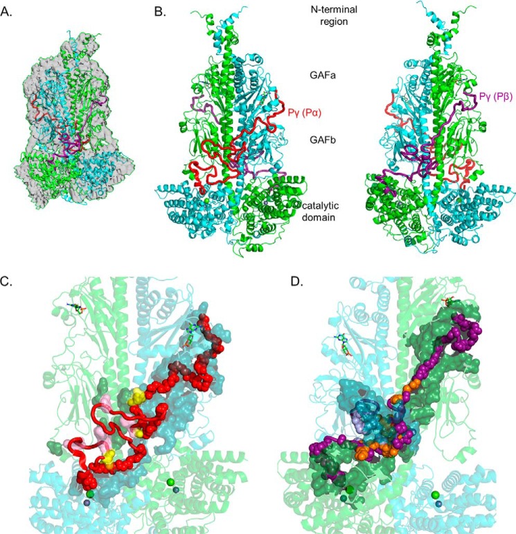
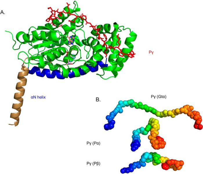
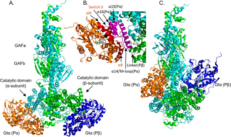
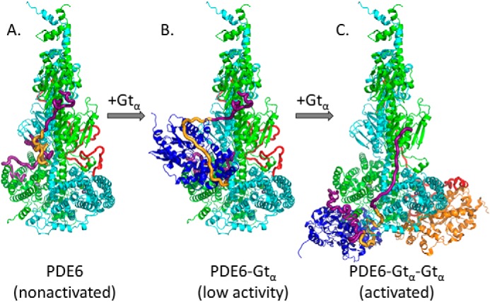
Photoreceptor phosphodiesterase 6 (PDE6) is the central effector of the visual excitation pathway in both rod and cone photoreceptors, and PDE6 mutations that alter PDE6 structure or regulation can result in several human retinal diseases. The rod PDE6 holoenzyme consists of two catalytic subunits (Pαβ) whose activity is suppressed in the dark by binding of two inhibitory γ-subunits (Pγ). Upon photoactivation of rhodopsin, the heterotrimeric G protein (transducin) is activated, resulting in binding of the activated transducin α-subunit (Gtα) to PDE6, displacement of Pγ from the PDE6 active site, and enzyme activation. Although the biochemistry of this pathway is understood, a lack of detailed structural information about the PDE6 activation mechanism hampers efforts to develop therapeutic interventions for managing PDE6-associated retinal diseases. To address this gap, here we used a cross-linking MS-based approach to create a model of the entire interaction surface of Pγ with the regulatory and catalytic domains of Pαβ in its nonactivated state. Following reconstitution of PDE6 and activated Gtα with liposomes and identification of cross-links between Gtα and PDE6 subunits, we determined that the PDE6-Gtα protein complex consists of two Gtα-binding sites per holoenzyme. Each Gtα interacts with the catalytic domains of both catalytic subunits and induces major changes in the interaction sites of the Pγ subunit with the catalytic subunits. These results provide the first structural model for the activated state of the transducin-PDE6 complex during visual excitation, enhancing our understanding of the molecular etiology of inherited retinal diseases.

Keywords: G protein; PDE6; allosteric regulation; integrative structural modeling; mass spectrometry (MS); phosphodiesterases; photoreceptor; phototransduction; protein cross-linking; transducin.


Conflict of interest statement

The authors declare that they have no conflicts of interest with the contents of this article

Figures

Figure 1.
Figure 1.
Integrative structural model of the PDE6 holoenzyme. The structural model of rod PDE6 holoenzyme (αβγγ) was determined by using the cryo-EM structure 6MZB (23) as a template and applying spatial restraints determined by chemical cross-linking of purified bovine rod PDE6 (Table 1 and Ref. 20). In the model, PDE6 subunits are colored as follows: α-subunit (Pα), cyan; β-subunit (Pβ), green; Pγ subunit primarily associated with α-subunit (Pγ(Pα)), red; and Pγ subunit primarily associated with β-subunit (Pγ(Pβ)), deep purple. A, superimposition of the template cryo-EM map (EMD-9297) with the cross-link–refined structural model of nonactivated PDE6 holoenzyme. B, asymmetric interactions of Pγ with the Pαβ catalytic dimer extending from the cGMP-binding GAFa domain to the GAFb domain and then crossing over to the catalytic domain to the site of inhibition of catalysis. Each Pγ subunit primarily interacts with one catalytic subunit. The two images are rotated 180°. C, interaction surface of the Pγ(Pα) subunit with the PDE6 catalytic dimer. Pγ(Pα) residues interacting with the catalytic dimer are shown as main-chain atom spheres: red, residues interacting with the α-subunit; pink, residues interacting with the β-subunit; and yellow, Pγ residues that interact with both catalytic subunits. Noninteracting Pγ(Pα) residues are shown as red loops and α-helix. The catalytic subunit interacting residues are shown as a surface representation (α-subunit, dark cyan; β-subunit, dark green). D, interaction surface of the Pγ(Pβ) subunit with Pαβ. The interaction surface of the Pγ(Pβ) subunit (180° rotation in C) is depicted in which the deep purple, light purple, and orange spheres represent interactions with the β-subunit, α-subunit, or both catalytic subunits, respectively.
Figure 2.
Figure 2.
Structural model of Gtα–GDP–AlF4 and its interaction with Pγ in solution. A, the structural model of Gtα was determined using the 1TAD crystal structure as the template (36) and refined with spatial restraints imposed from cross-linking results in the absence or presence of Pγ (Table 2). Structural elements that were unchanged in the cross-link–refined model are represented in green, with the conformational change of the αN helix shown in brown (for the crystal structure) and blue (for the cross-link modified solution structure). Also shown is the docked structure of Pγ (red) with Gtα–GDP–AlF4 based on the observed cross-linking results when Gtα associated with lipobeads was incubated with a 2-fold molar excess of Pγ. Note that no significant changes in Gtα conformation were observed upon Pγ binding. B, a comparison of the conformation of the central region of Pγ (residues 24–44, depicted as a gradient from blue to red spheres) when bound to Gtα or to the PDE6 catalytic subunits.
Figure 3.
Figure 3.
Model of Gtα–GDP–AlF4 docked to the Pαβ catalytic dimer. PDE6 holoenzyme and Gtα–GDP–AlF4 bound to lipobeads (see “Experimental procedures”) were exposed to chemical cross-linkers, and the identified cross-linked peptides between Gtα and PDE6 subunits (Table 3) were then used as spatial restraints for integrative structural modeling. Two predominant clusters of models of the Gtα–Pαβ complex were generated, one with Gtα docked to the two catalytic domains (with distance violations for Gtα24-Pα330/Pβ328, Gtα9-Pα442, and Gtα9-Pβ440) and the other with Gtα docked to the GAFb domains (with distance violations for Gtα10-Pα854, Gtα17-Pα551, and Gtα128-Pα807/Pβ817). Because of insufficient cross-links for Pγ in the activated complex, the inhibitory subunit is not shown. A, structural model of association of Gtα–GDP–AlF4 to the α-subunit (Gtα(Pα), orange) and to the β-subunit (Gtα(Pβ), blue) catalytic domains. B, detailed view of the Gtα GTPase subdomain interface with the α-subunit catalytic domain, with the interaction surface of Gtα colored red and the α- and β-subunit interacting residues colored magenta and brown, respectively. The black sphere indicates Gtα Gln200. C, alternate docking of Gtα to the GAFb domains of the Pαβ catalytic dimer (with the same orientation as in A).
Figure 4.
Figure 4.
Proposed model for the activation of PDE6 by transducin during visual excitation. A, in the dark-adapted condition, the PDE6 holoenzyme is inhibited by its Pγ subunits occluding the enzyme active site (Fig. 1B, rotated 90°). B, the first light-activated Gtα subunit is proposed to initially bind to the GAFb docking site (see Fig. 3C) without causing significant catalytic activation of PDE6 (21). The Pγ subunit was docked to this complex using the following information: (a) the central region of Pγ (gold) was docked using the cross-links obtained for the Gtα–Pγ complex (Table 2) in conjunction with the cross-links used to dock Gtα to the GAFb domain (Table 3); (b) lacking cross-linking data for the N-terminal region of Pγ in the activated complex, this region of Pγ (purple) relied on PDE6 holoenzyme cross-links, and thus its topology only differs from Fig. 1 to the extent needed to accommodate cross-link spatial restraints imposed by the Pγ central region; and (c) in the absence of Pγ cross-links for its C-terminal region in the activated complex, we modeled this region of Pγ (purple) interacting with Gtα using the crystal structure of Pγ (residues 50–87) bound to a chimeric G protein (PDB code 1FQJ) (11). C, upon binding of a second Gtα, PDE6 becomes fully activated as both Gtα subunits dock to the catalytic domains and displace the C-terminal region of Pγ from the enzyme active sites. To accommodate the binding of the central region of Pγ to the helical face (Table 2) and the C-terminal region of Pγ to the GTPase face of Gtα (Table 3 and Ref. 11), a major displacement of the N-terminal Pγ residues from the GAFa domains must occur.

References

    1. Arshavsky V. Y., and Burns M. E. (2012) Photoreceptor signaling: supporting vision across a wide range of light intensities. J. Biol. Chem. 287, 1620–1626 10.1074/jbc.R111.305243 - DOI - PMC - PubMed
    1. Bender A. T., and Beavo J. A. (2006) Cyclic nucleotide phosphodiesterases: molecular regulation to clinical use. Pharmacol. Rev. 58, 488–520 10.1124/pr.58.3.5 - DOI - PubMed
    1. Francis S. H., Blount M. A., and Corbin J. D. (2011) Mammalian cyclic nucleotide phosphodiesterases: molecular mechanisms and physiological functions. Physiol. Rev. 91, 651–690 10.1152/physrev.00030.2010 - DOI - PubMed
    1. Zoraghi R., Corbin J. D., and Francis S. H. (2004) Properties and functions of GAF domains in cyclic nucleotide phosphodiesterases and other proteins. Mol. Pharmacol. 65, 267–278 10.1124/mol.65.2.267 - DOI - PubMed
    1. Cote R. H. (2006) Photoreceptor phosphodiesterase (PDE6): a G-protein-activated PDE regulating visual excitation in rod and cone photoreceptor cells. In Cyclic Nucleotide Phosphodiesterases in Health and Disease (Beavo J. A., Francis S. H., and Houslay M. D., eds) pp. 165–193, CRC Press, Boca Raton, FL

Publication types