Comparative Study
. 2019 Nov 5;9(1):16310.
doi: 10.1038/s41598-019-52213-6.

Proof-of-Concept Study of Drug Brain Permeability Between in Vivo Human Brain and an in Vitro iPSCs-Human Blood-Brain Barrier Model

Affiliations
Comparative Study

Proof-of-Concept Study of Drug Brain Permeability Between in Vivo Human Brain and an in Vitro iPSCs-Human Blood-Brain Barrier Model

Gwenaëlle Le Roux et al. Sci Rep. .

Abstract

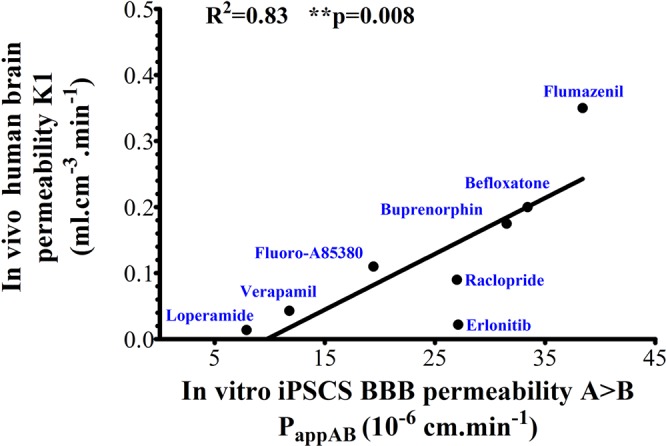
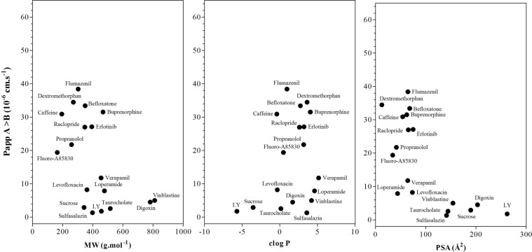
The development of effective central nervous system (CNS) drugs has been hampered by the lack of robust strategies to mimic the blood-brain barrier (BBB) and cerebrovascular impairments in vitro. Recent technological advancements in BBB modeling using induced pluripotent stem cells (iPSCs) allowed to overcome some of these obstacles, nonetheless the pertinence for their use in drug permeation study remains to be established. This mandatory information requires a cross comparison of in vitro and in vivo pharmacokinetic data in the same species to avoid failure in late clinical drug development. Here, we measured the BBB permeabilities of 8 clinical positron emission tomography (PET) radioligands with known pharmacokinetic parameters in human brain in vivo with a newly developed in vitro iPSC-based human BBB (iPSC-hBBB) model. Our findings showed a good correlation between in vitro and in vivo drug brain permeability (R2 = 0.83; P = 0.008) which contrasted with the limited correlation between in vitro apparent permeability for a set of 18 CNS/non-CNS compounds using the in vitro iPSCs-hBBB model and drug physicochemical properties. Our data suggest that the iPSC-hBBB model can be integrated in a flow scheme of CNS drug screening and potentially used to study species differences in BBB permeation.


Conflict of interest statement

The authors declare no competing interests.

Figures

Figure 1
Figure 1
Establishment and characterization of iPSCs. (A) Representative analysis of TRA1-60 and SSEA-4 expression for each iPSC cell line by flow cytometry. TRA1-60 is compared with unlabeled cells and SSE-A4 is compared with the appropriate mouse IgG3 control. Control: green line; test: purple line. Samples of somatic control cells (Caco-2 epithelial cells for TRA1-60 and hBMEC for SSEA-4) were tested in parallel (respectively). Flow cytometry analysis of the iPSCs lines were performed in exponential growth phase. (B) Gene expression analysis of pluripotency markers in both iPSC and in fibroblast control by semi-quantitative PCR. (C) Ability of the iPSC-SP cell line to generate all three germ cell layer components within teratomas (endoderm – line 1, mesoderm – line 2 and ectoderm – line 3) was evaluated. Whole sections of teratoma were performed after 5 to 10 weeks of growth and stained with hematoxylin and eosin. Scale bare = 50 µm.
Figure 2
Figure 2
Differentiation of iPSC-hBECs. (A) Representative phase contrast images of iPSCs in cell culture at day 5 and after differentiation. By day 8, cells exhibited brain endothelial cell morphology in the presence of puromycin. Black scale bare, 500 µm. White scale bare, 50 µm. (B) ZO-1 and claudin-5 fluorescence intensity demonstrated that the differentiated cells exhibited brain endothelial characteristics. Scale bare, 50 µm. (C) Typical profile of transporter/receptor gene expression. The level of the expression of mRNA of junctional proteins like claudin-5 and ZO-1, transporters like ABCC1, ABCG2 and LRP1 in iPSC-Human BECs was comparable to that observed in our two primary hBECs samples.
Figure 3
Figure 3
Low paracellular transport of sucrose and high permeability of propranolol in the in vitro iPSC-based human BBB model. (A,C) Each column represents the average of the apparent permeability (mean ± SD) from apical to basal compartment obtained on independent iPSC-BBB in vitro models (7 to 8 experiments). (B,D) each column represents the average of the apparent permeability from apical to basal compartment obtained in the two cell lines (mean ± SEM).
Figure 4
Figure 4
Assessment of the bidirectional transport of the P-gp transporter substrate vinblastine in the iPSC-hBBB models. (A) ER of iPSC-hBBB models from different iPSC batches. (B) ER mean ratio in iPSC-hBBB models from at least 6 identical iPSC showing the ER intra-variability (mean ± SEM). (C) Effect of CsA on the vinblastine ER (mean ± SD).
Figure 5
Figure 5
Clinical validation of iPSC- hBBB Blood-Brain Barrier model. Linear regression analysis shows a positive correlation between apparent permeability in human brain (K1) and in vitro apparent permeability in the iPSC-hBBB model. Spearman tests were performed and *P < 0.05 indicates y significant correlation.
Figure 6
Figure 6
iPSC- hBBB Blood-Brain Barrier model permeability characterization. Apparent permeability values of marketed CNS and non-CNS drugs from the in vitro iPSC-hBBB model and relationship with their molecular weight (MW), cLogP, and surface polar area (PSA). Data are the average of two or three cell monolayers for each tested compound.
Figure 7
Figure 7
Species differences in blood-brain barrier transport. (A) Species correlation of apparent permeability in blood-brain barrier transport. (B) Species correlation of efflux ratio between in vitro primary rat cell based BBB model and in vitro iPSC-hBBB model. The dotted lines represent limits of ER.

References

    1. Abbott NJ, Patabendige AAK, Dolman DEM, Yusof SR, Begley DJ. Structure and function of the blood-brain barrier. Neurobiol. Dis. 2010;37:13–25. doi: 10.1016/j.nbd.2009.07.030. - DOI - PubMed
    1. Omidi Y, Barar J. Impacts of blood-brain barrier in drug delivery and targeting of brain tumors. Bioimpacts. 2012;2:5–22. - PMC - PubMed
    1. Dong X. Current Strategies for Brain Drug Delivery. Theranostics. 2018;8:1481–1493. doi: 10.7150/thno.21254. - DOI - PMC - PubMed
    1. Hurko O, Ryan JL. Translational Research in Central Nervous System Drug Discovery. NeuroRx. 2005;2:671–682. doi: 10.1602/neurorx.2.4.671. - DOI - PMC - PubMed
    1. Josserand V, et al. Evaluation of drug penetration into the brain: a double study by in vivo imaging with positron emission tomography and using an in vitro model of the human blood-brain barrier. J. Pharmacol. Exp. Ther. 2006;316:79–86. doi: 10.1124/jpet.105.089102. - DOI - PubMed

Publication types