. 2019 Nov 5;9(1):16039.
doi: 10.1038/s41598-019-52366-4.

A Novel Semiconductor-Based Flow Cytometer with Enhanced Light-Scatter Sensitivity for the Analysis of Biological Nanoparticles

Affiliations

A Novel Semiconductor-Based Flow Cytometer with Enhanced Light-Scatter Sensitivity for the Analysis of Biological Nanoparticles

George C Brittain 4th et al. Sci Rep. .

Abstract

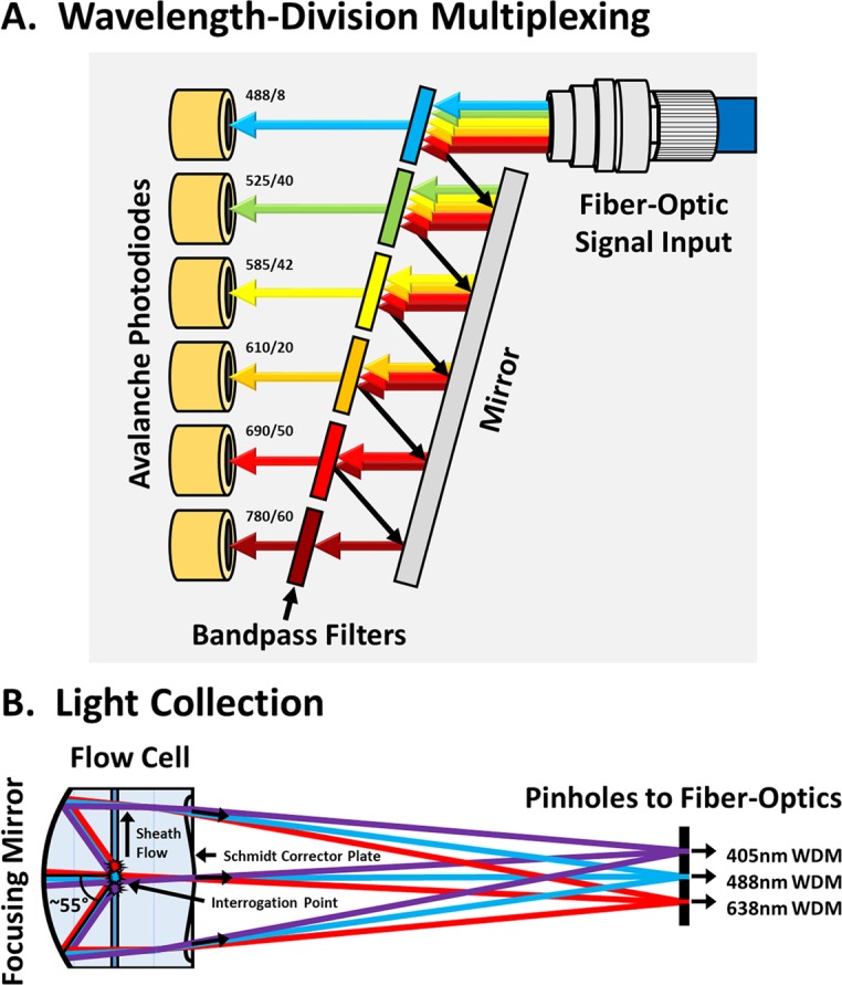
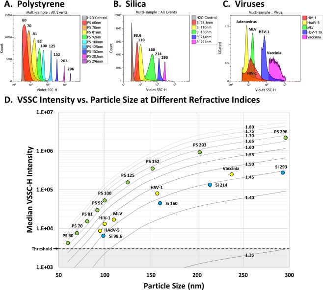
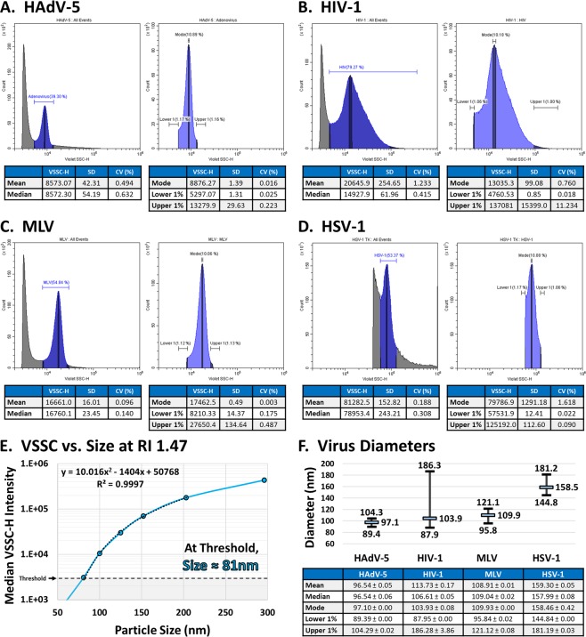
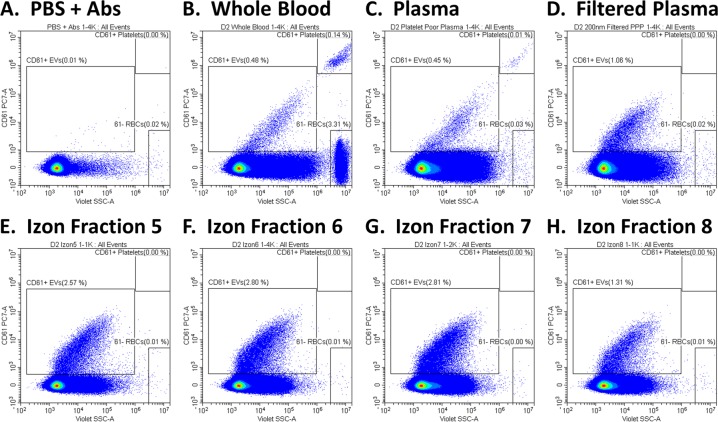
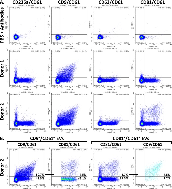
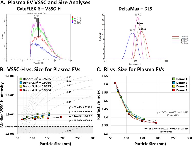
The CytoFLEX is a novel semiconductor-based flow cytometer that utilizes avalanche photodiodes, wavelength-division multiplexing, enhanced optics, and diode lasers to maximize light capture and minimize optical and electronic noise. Due to an increasing interest in the use of extracellular vesicles (EVs) as disease biomarkers, and the growing desire to use flow cytometry for the analyses of biological nanoparticles, we assessed the light-scatter sensitivity of the CytoFLEX for small-particle detection. We found that the CytoFLEX can fully resolve 70 nm polystyrene and 98.6 nm silica beads by violet side scatter (VSSC). We further analyzed the detection limit for biological nanoparticles, including viruses and EVs, and show that the CytoFLEX can detect viruses down to 81 nm and EVs at least as small as 65 nm. Moreover, we could immunophenotype EV surface antigens, including directly in blood and plasma, demonstrating the double labeling of platelet EVs with CD61 and CD9, as well as triple labeling with CD81 for an EV subpopulation in one donor. In order to assess the refractive indices (RIs) of the viruses and EVs, we devised a new method to inversely calculate the RIs using the intensity vs. size data together with Mie-theory scatter efficiencies scaled to reference-particle measurements. Each of the viruses tested had an equivalent RI, approximately 1.47 at 405 nm, which suggests that flow cytometry can be more broadly used to easily determine virus sizes. We also found that the RIs of EVs increase as the particle diameters decrease below 150 nm, increasing from 1.37 for 200 nm EVs up to 1.61 for 65 nm EVs, expanding the lower range of EVs that can be detected by light scatter. Overall, we demonstrate that the CytoFLEX has an unprecedented level of sensitivity compared to conventional flow cytometers. Accordingly, the CytoFLEX can be of great benefit to virology and EV research, and will help to expand the use of flow cytometry for minimally invasive liquid biopsies by allowing for the direct analysis of antigen expression on biological nanoparticles within patient samples, including blood, plasma, urine and bronchoalveolar lavages.


Conflict of interest statement

G.C.B., E.M. and S.G. are employees of Beckman Coulter, Inc. Y.Q.C. was the VP of Research and CTO of Beckman Coulter, Inc. M.-A.L. is the CEO and V.A.T. is the CSO of ViroFlow Technologies, Inc.

Figures

Figure 1
Figure 1
Optical Innovations in the CytoFLEX. (A) Wavelength-division multiplexing is a method for parsing ranges of light wavelengths, adapted from fiber-optics technology used in the telecommunications industry. Input light from the fiber-optic cable is sequentially reflected by bandpass filters until the particular wavelength range encounters a permissive filter that allows the light to pass through to its associated APD. This design minimizes the loss of light, as occurs with dichroic mirrors. (B) The catadioptric flow-cell design maximizes light collection. Approximately 110° of side-scatter and fluorescent light is focused by a plano-concave mirror on the back of the flow cell. The light path is then shaped by a lens designed similar to a Schmidt corrector plate, which directs light originating from the different lasers to their respective fiber-optic pinholes, while also minimizing the cross-mixing of light.
Figure 2
Figure 2
VSSC Sensitivity. (A) The detection of 60–296 nm PS particles by VSSC. The CytoFLEX can detect as small as 60 nm PS nanoparticles, and can resolve as small as 70 nm. (B) The detection of 98.6–293 nm Si particles by VSSC. 98.6 nm Si particles with a RI of 1.44 can be fully resolved by VSSC. (C) VSSC detection of viruses. Unlabeled 95 nm HAdV-5, 100 nm HIV-1, and 110 nm MLV can be fully resolved by light scatter on the CytoFLEX. (D) A contour plot prepared using Mie-theory RI curves scaled to the CytoFLEX VSSC intensities. The data from (A–C) were overlaid on the plot to verify the accuracy of the scaling. All samples were collected in triplicate and these data represent the population means. VSSC gain = 400; VSSC-H threshold = 3000. The VSSC-H threshold for HSV-1 and Vaccinia was 40 K and 100 K, respectively.
Figure 3
Figure 3
Sizing Viruses by Flow Cytometry. (A–D) VSSC-H population statistics for (A) HAdV-5, (B) HIV-1, (C) MLV, and (D) HSV-1. Each virus was read in triplicate and the data represent the average. (E) Fitting a VSSC-H Intensity vs. Size curve for RI 1.47 at 405 nm in order to calculate virus sizes. (F) The calculated size characteristics for each virus in (A–D). In the chart above, the calculated peak diameter is displayed along with the size range. The mean and median sizes are also included in the table below, ±the SEM.
Figure 4
Figure 4
Preparation of Plasma EVs. (A) PBS + antibody cocktail as a control to demonstrate the absence of fluorescent-antibody aggregates. (B) CD61+ EVs and platelets in whole blood. (C) CD61+ EVs in PPP, with RBCs eliminated and platelets depleted. (D) CD61+ EVs in PPP filtered through a 0.2 μm filter, with platelets eliminated. (E–H) CD61+ EVs in the 5th through 8th 200 μL fraction from 0.2 μm-filtered PPP passed through an Izon qEV column. Serial dilutions were performed and all samples were acquired in triplicate. VSSC gain = 400; VSSC-H threshold = 3000.
Figure 5
Figure 5
Tetraspanin Expression on the CD61+ plasma EVs. (A) CD61+ plasma EVs from human donors were tested for their expression of CD9, CD63 and CD81. The CD61+ EVs were around 50% CD9+, while CD63 and CD81 expression was mostly absent. 8.7% of the CD61+ EVs from Donor 2 were CD81+. The PBS + antibody control demonstrates the lack of fluorescent-antibody aggregates. (B) Further analysis of the CD81+ EVs revealed that these were predominantly CD9, CD81 and CD61 triple positive. This is reciprocally demonstrated by analyzing the CD81 expression on CD9+ CD61+ EVs, and the CD9 expression on CD81+ CD61+ EVS. In total, 7.5% of the CD61 EVs from Donor 2 were triple positive. The percentages in bold are in reference to the overall CD61+ EV population. These samples were serially diluted and acquired in triplicate. VSSC gain = 400; VSSC-H threshold = 3000.
Figure 6
Figure 6
Analysis of the VSSC Sensitivity for EV Detection. (A) Analysis of the purified EV fractions by VSSC intensity and DLS sizing. The overlay plots demonstrate the differential VSSC intensities and DLS sizes for representative measurements of the EV fractions from Donor 2. (B) Overlay of the average VSSC intensity vs. DLS size measurements for each EV fraction on the scaled Mie-theory RI curves prepared in Fig. 2. The sample distributions are almost perfectly linear, with R2 values between 0.930 and 0.997 relative to linear trendlines. (C) The RIs of the EV fractions increase progressively as their sizes decrease. All samples were serially diluted and the VSSC-H intensity measurements were acquired in triplicate or quadruplicate, while 100 DLS measurements were acquired per sample. VSSC gain = 400; VSSC-H threshold = 3000.

References

    1. Welsh JA, Holloway JA, Wilkinson JS, Englyst NA. Extracellular vesicle flow cytometry analysis and standardization. Front. Cell Dev. Biol. 2017;5:78. doi: 10.3389/fcell.2017.00078. - DOI - PMC - PubMed
    1. Willms E, Cabañas C, Mäger I, Wood MJA, Vader P. Extracellular vesicle heterogeneity: subpopulations, isolation techniques, and diverse functions in cancer progression. Front. Immunol. 2018;9:738. doi: 10.3389/fimmu.2018.00738. - DOI - PMC - PubMed
    1. Stoner SA, et al. High sensitivity flow cytometry of membrane vesicles. Cytometry A. 2016;89(2):196–206. doi: 10.1002/cyto.a.22787. - DOI - PubMed
    1. Van der Pol E, Böing AN, Harrison P, Sturk A, Nieuwland R. Classification, functions, and clinical relevance of extracellular vesicles. Pharmacol. Rev. 2012;64:676–705. doi: 10.1124/pr.112.005983. - DOI - PubMed
    1. Sodar BW, et al. Low-density lipoprotein mimics blood plasma-derived exosomes and microvesicles during isolation and detection. Sci. Rep. 2016;6:24316. doi: 10.1038/srep24316. - DOI - PMC - PubMed

Publication types