. 2019 Nov 5;9(1):16046.
doi: 10.1038/s41598-019-52458-1.

Dynamic Quantitative Iodine Myocardial Perfusion Imaging with Dual-Layer CT using a Porcine Model

Affiliations

Dynamic Quantitative Iodine Myocardial Perfusion Imaging with Dual-Layer CT using a Porcine Model

Kai Scherer et al. Sci Rep. .

Abstract

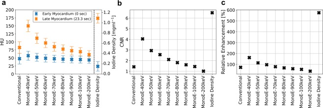
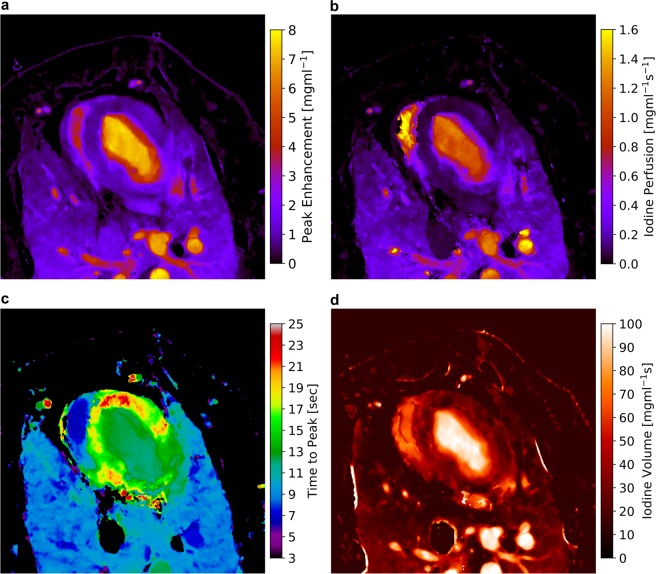
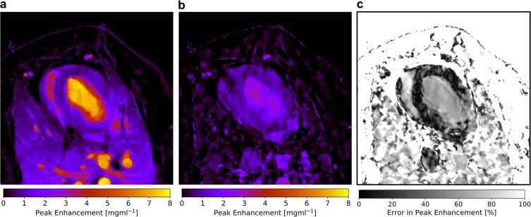
Ischemic heart disease is the globally leading cause of death. When using coronary CT angiography, the functional hemodynamics within the myocardium remain uncertain. In this study myocardial CT perfusion imaging using iodine contrast agent demonstrated to strongly improve the assessment of myocardial disorders. However, a retrieval of such dynamics using Hounsfield units from conventional CT poses concerns with respect to beam-hardening effects and low contrast-to-noise ratio (CNR). Dual-energy CT offers novel approaches to overcome aforementioned limitations. Quantitative peak enhancement, perfusion, time to peak and iodine volume measurements inside the myocardium were determined resulting in 0.92 mg/ml, 0.085 mg/ml/s 17.12 s and 29.89 mg/ml*s, respectively. We report on the first extensive quantitative and iodine-based analysis of myocardial dynamics in a healthy porcine model using a dual-layer spectral CT. We further elucidate on the potential of reducing the radiation dose from 135 to 18 mGy and the contrast agent volume from 60 to 30 mL by presenting a two-shot acquisition approach and measuring iodine concentrations in the myocardium in-vivo down to 1 mg/ml, respectively. We believe that dynamic quantitative iodine perfusion imaging may be a highly sensitive tool for the precise functional assessment and monitoring of early myocardial ischemia.


Conflict of interest statement

The authors declare no competing interests.

Figures

Figure 1
Figure 1
(a) Conventional and mono-energetic Hounsfield units and iodine density of the early (blue points) and late myocardium (orange). The early stage is measured at second zero without iodine enhancement, the late myocardial stage is depicted at second 23.3 with maximal contrast agent enrichment. Error bars correspond to the standard deviation found within a ROI of 50 mm2 of the respective image channels. (b) Temporal contrast-to-noise and (c) relative signal enhancement of the myocardium during contrast uptake derived from (a), demonstrating a superior depiction of the myocardial opacification within the iodine density imaging channel.
Figure 2
Figure 2
(a) Measured transversal quantitative iodine density slices of the porcine thorax showing a subsequent opacification within the right (RV) and left heart ventricle (LV) as well as the myocardium (Myo). (b) Iodine density values vs. scan time as obtained from the dynamic dual-energy perfusion CT for an exemplary pixel for various region of interest as indicated by yellow boxes in (a), which were selected by an experienced radiologist. A high accordance of data and fit was obtained using a gamma variate fit model. Note that only the first pass of contrast agent was considered, and a recirculation of blood was correspondingly neglected for the fit. (c) Transversal quantitative iodine density slices of the porcine thorax obtained from the model being in good agreement with the measured data as shown in (a). Note that distinctive discrepancies for late scan times arise from the neglection of recirculation.
Figure 3
Figure 3
Absolute and quantitative iodine-based analysis of myocardial dynamics: (a) peak enhancement, (b) perfusion, (c) time to peak and (d) volume maps of the porcine thorax, derived from the gamma variate fit model using the slope method. The peak enhancement indicates the maximal increase in iodine density, the perfusion relates to the highest temporally gradient in influx of iodine, the time to peak indicates the point in time when maximal peak enhancement is reached, and the iodine volume is related to uptake/storing/flush out behaviour within the respective structures. Note that areas, where the gamma variate fit failed (for instance in the cava inferior) or areas where the iodine density remained below 0.55 mg/ml during the scan are color-coded black.
Figure 4
Figure 4
(a) Quantitative iodine-based peak enhancement of the porcine thorax as derived from the dynamics analysis based on 15 scans. (b) Quantitative iodine-based density peak enhancement of the porcine thorax as derived from a semi-static two-shot approach during myocardial contrast saturation. Notice that arterial and ventricular structures are strongly underestimated in comparison to (a). (c) Relative error in the determination of the peak enhancement of (b) in comparison to the ground truth (a). Within the myocardium the deviation is quite homogeneously patterned and accounts to values in the range of 5–15%.

References

    1. Mozaffarian D, et al. Heart Disease and Stroke Statistics - 2015 Update. A Report from the American Heart Association. Circulation. 2015;131:29–322. - PubMed
    1. Finegold J, Asaria P, Francis D. Mortality from ischaemic heart disease by country, region, and age: statistics from World Health Organisation and United Nations. Int. J. Cardiol. 2013;168:934–945. doi: 10.1016/j.ijcard.2012.10.046. - DOI - PMC - PubMed
    1. Rossi A, et al. Diagnostic performance of hyperaemic myocardial blood flow index obtained by dynamic computed tomography: does it predict functionally significant coronary lesions? Eur. Heart J. Cardiovasc. Imaging. 2014;15:85–94. doi: 10.1093/ehjci/jet133. - DOI - PubMed
    1. Bittencourt M, et al. Clinical outcomes after evaluation of stable chest pain by coronary computed tomographic angiography versus usual care: a meta-analysis. Circ. Cardiovasc. Imaging. 2016;9:e004419. - PubMed
    1. Joergensen M, et al. Functional testing or coronary computed tomography angiography in patients with stable coronary artery disease. J. Am. Coll. Cardiol. 2017;69:1761–1770. doi: 10.1016/j.jacc.2017.01.046. - DOI - PubMed

Publication types