. 2019 Dec;7(4):326-341.
doi: 10.1002/iid3.277. Epub 2019 Nov 5.

Ischemic stroke alters immune cell niche and chemokine profile in mice independent of spontaneous bacterial infection

Affiliations

Ischemic stroke alters immune cell niche and chemokine profile in mice independent of spontaneous bacterial infection

Breanne Y Farris et al. Immun Inflamm Dis. 2019 Dec.

Abstract

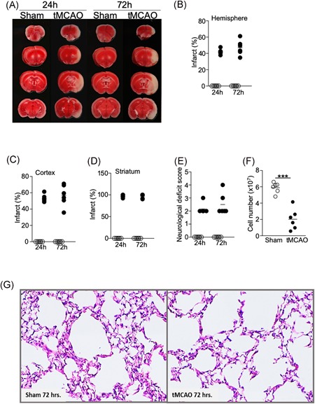
Introduction: Stroke-associated pneumonia (SAP) is a major cause of mortality in patients who have suffered from severe ischemic stroke. Although multifactorial in nature, stroke-induced immunosuppression plays a key role in the development of SAP. Previous studies using a murine model of transient middle cerebral artery occlusion (tMCAO) have shown that focal ischemic stroke induction results in functional defects of lymphocytes in the spleen, thymus, and peripheral blood, leading to spontaneous bacterial infection in the lungs without inoculation. However, how ischemic stroke alters immune cell niche and the expression of cytokines and chemokines in the lungs has not been fully characterized.

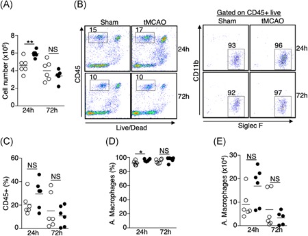
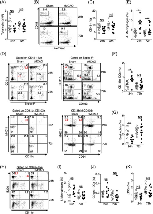
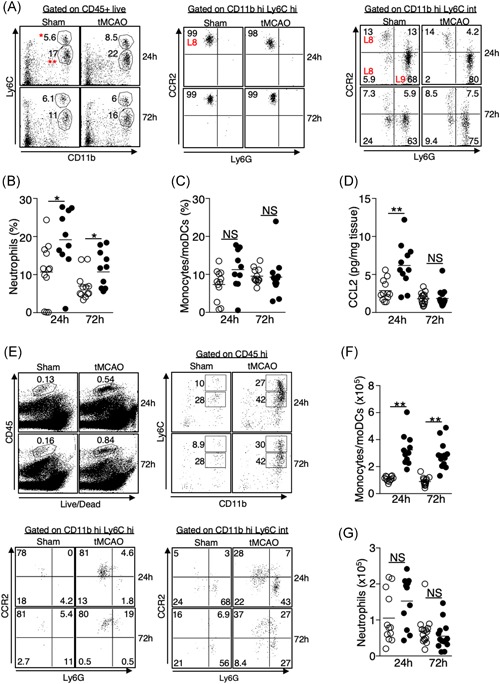
Methods: Ischemic stroke was induced in mice by tMCAO. Immune cell profiles in the brain and the lungs at 24- and 72-hour time points were compared by flow cytometric analysis. Cytokine and chemokine expression in the lungs were determined by multiplex bead arrays. Tissue damage and bacterial burden in the lungs following tMCAO were evaluated.

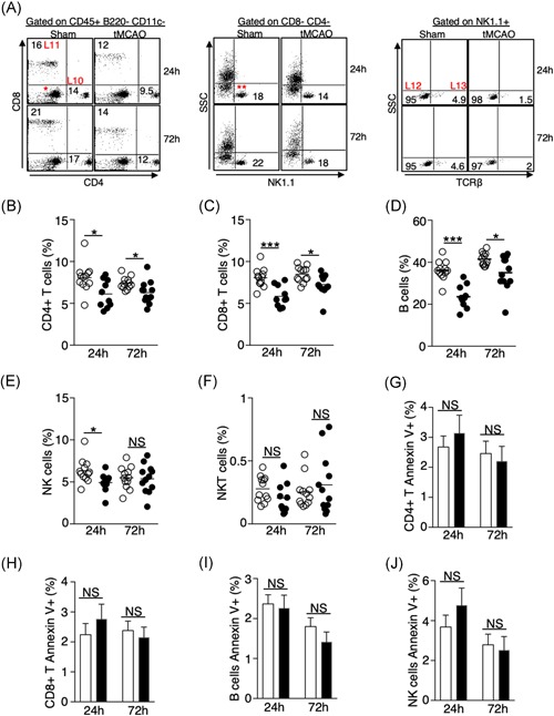
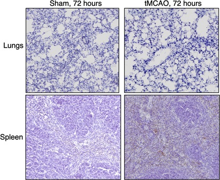
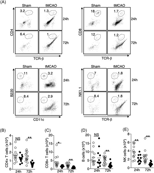
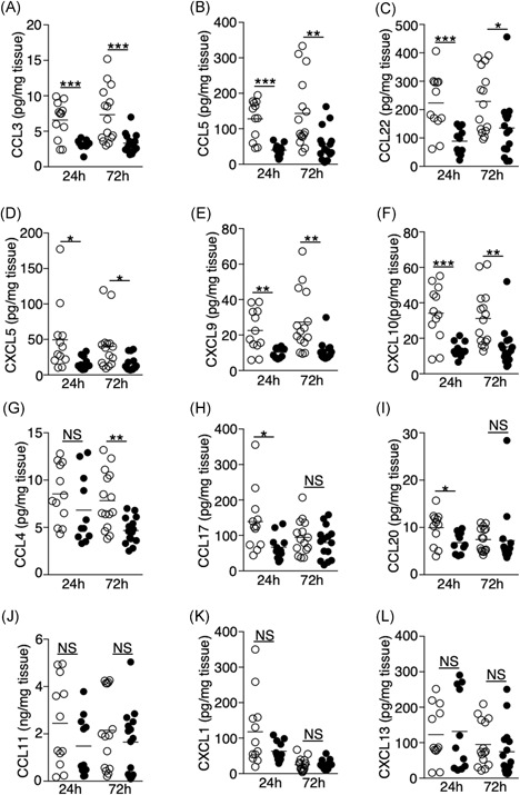
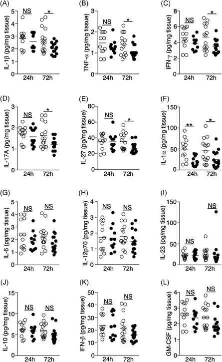
Results: Ischemic stroke increases the percentage of alveolar macrophages, neutrophils, and CD11b+ dendritic cells, but reduces the percentage of CD4+ T cells, CD8+ T cells, B cells, natural killer cells, and eosinophils in the lungs. The alteration of immune cell niche in the lungs coincides with a significant reduction in the levels of multiple chemokines in the lungs, including CCL3, CCL4, CCL5, CCL17, CCL20, CCL22, CXCL5, CXCL9, and CXCL10. Spontaneous bacterial infection and tissue damage following tMCAO, however, were not observed.

Conclusion: This is the first report to demonstrate a significant reduction of lymphocytes and multiple proinflammatory chemokines in the lungs following ischemic stroke in mice. These findings suggest that ischemic stroke directly impacts pulmonary immunity.

Keywords: chemokines; pulmonary immunity; stroke.


Conflict of interest statement

The authors declare that there are no conflict of interests.

Figures

Figure 1
Figure 1
Severe ischemic stroke in C57BL/6J mice does not cause spontaneous pneumonia. Brain, spleen, and lung tissues were analyzed 24 and 72 hours following tMCAO or sham operation. A, Representative images showing ipsilateral brain infarcts following tMCAO but not sham operation by TTC staining. B‐D, Percentage of infarcts within the ipsilateral hemisphere (B), cortex (C), and corpus striatum (D) following tMCAO (filled circle) or sham controls (open circle) quantified by Image J. E, Neurological deficit scores of the mice 24 and 72 hours following tMCAO (filled circle) or sham controls (open circle). See Section 2 for score definition. F, Cell number from the spleens of mice 72 hours following tMCAO (filled circle) or sham controls (open circle). G, Representative images from H&E staining of lung tissues 72 hours following tMCAO (right) or sham operation (left). Images from all animals are shown in Figure S1. Data shown are combined results from two independent experiments with n = 6 animals per group (sham 24 hours, tMCAO 24 hours, sham 72 hours, and tMCAO 72 hours). ***P < .001. H&E, hematoxylin and eosin; tMCAO, transient middle cerebral artery occlusion; TTC, Triphenyltetrazolium chloride
Figure 2
Figure 2
Increase in the number of alveolar macrophages in the BALF 24 hours postischemic stroke. A, Total number of cells recovered from BALF 24 and 72 hours following tMCAO (filled circle) or sham operation (open circle). B, Cellular compositions of BALF 24 and 72 hours following tMCAO. B, Representative plots showing percentage of CD45+ cells (left) and alveolar macrophages (right), which are defined as CD45+ Siglec F+ CD11b−. C‐E, Graphs showing percentage of CD45+ cells (C); percentage (D) and number (E) of alveolar macrophages of individual animals described in (B). tMCAO (filled circle) and sham operation (open circle). Data shown are combined results from two independent experiments with n = 6 animals per group (sham 24 hours; tMCAO 24 hours; sham 72 hours; tMCAO 72 hours). *P < .05; **P < .01. BALF, bronchoalveolar lavage fluid; NS, not statistically different; tMCAO, transient middle cerebral artery occlusion
Figure 3
Figure 3
Alterations in the resident innate immune cell niche in the lungs following ischemic stroke. Lung tissues were excised 24 and 72 hours following tMCAO (filled circle) or sham operation (open circle), resident innate immune cells in the lungs were analyzed by flow cytometry, defined by surface markers listed on Table 1. A, Graph showing total number of cells in the lungs of individual animals. B, Representative plots showing percentage of CD45+ cells. C, Graph showing percentage of CD45+ cells in the lungs of individual animals. D, Representative plots showing the identification of alveolar macrophages (L1), eosinophils (L2), CD103+ DCs (L3), CD11b+ DCs (L4), and interstitial macrophages (L5). CD103+ DCs were first gated on CD103+ CD11b− cells (*). CD11b+ DCs and interstitial macrophages were first gated on CD11b hi CD103− cells (**). E‐G, Graphs showing percentage of alveolar macrophages (E), CD11b+ DCs (F), and eosinophils (G) of individual animals. H, Representative plots showing that identification of pDCs (L6), and B cells (L7, discussed in Figure 5). I‐K, Graphs showing percentage of interstitial macrophages (I), CD103+ DCs (J), and pDCs (K) of individual animals. Data shown are combined results from three independent experiments with n = 12 animals per group (sham 24 hours; tMCAO 24 hours; sham 72 hours; tMCAO 72 hours). **P < .01; ***P < .001. NS, not statistically different; pDC, plasmacytoid dendritic cell; tMCAO, transient middle cerebral artery occlusion
Figure 4
Figure 4
Increased infiltration of neutrophils but not monocytes to the lungs following ischemic stroke despite an elevation of CCL2. A‐C, Lung tissues were excised 24 and 72 hours following tMCAO or sham operation. Monocytes/moDCs and neutrophils in the lungs were analyzed by flow cytometry, defined by surface markers listed on Table 1. A, Representative plots showing the identification of monocytes/moDCs (L8) and neutrophils (L9) in the lungs. Monocytes/moDCs were defined as CD45+ Ly6C hi (*) CCR2 hi (middle) and Ly6C intermediate (**) CCR2+/− (right). Neutrophils were defined as CD45+ Ly6C intermediate (**) Ly6G+ (right). B‐C, Graphs showing percentage of neutrophils (B) and monocytes/moDCs (C) of individual animals 24 and 72 hours following tMCAO (filled circle) or sham operation (open circle). D, Lung tissues were homogenized 24 and 72 hours following tMCAO (filled circle) or sham operation (open circle), level of CCL2 was determined by multiplex bead array. E‐G, Brain tissues were excised 24 and 72 hours following tMCAO or sham operation, monocytes/moDCs and neutrophils in the brains were analyzed by flow cytometry. E, Representative plots showing the identification of monocytes/moDCs and neutrophils in the brains, which were defined as in (A). F,G, Graphs showing number of monocytes/moDCs (F) and neutrophils (G) of individual animals 24 and 72 hours following tMCAO (filled circle) or sham operation (open circle). Data shown are combined results from three independent experiments with n = 12 animals per group (sham 24 hours; tMCAO 24 hours; sham 72 hours; tMCAO 72 hours). *P < .05; **P < .01. moDC, monocyte‐derived dendritic cell; NS, not statistically different; tMCAO, transient middle cerebral artery occlusion
Figure 5
Figure 5
Ischemic stroke leads to a significant loss of lymphocytes in the lungs independent of apoptosis. A, Representative plots showing the identification of lymphocytes in the lungs by surface markers listed on Table 1. CD4+ (L10) and CD8+ (L11) T cells were first gated on CD45+ B220− CD11c− cells shown in Figure 3H. NK cells (L12) and NKT cells (L13) were gated on CD4− CD8− cells (*), then further gated on NK1.1+ cells (**). Representative plots for the identification of B cells shown in Figure 3H. B‐F, Graphs showing percentage of CD4+ T cells (B), CD8+ T cells (C), B cells (D), NK cells (E), and NKT cells (F) of individual animals 24 and 72 hours following tMCAO (filled circle) or sham operation (open circle). G‐H, Graphs showing percentage of annexin‐V+ CD4+ T cells (G), CD8+ T cells (H), B cells (I), and NK cells (J) 24 and 72 hours following tMCAO (filled bar) or sham operation (open bar). Data shown are combined results from three independent experiments with n = 12 animals per group (sham 24 hours, tMCAO 24 hours, sham 72 hours, tMCAO 72 hours). *P < .05; ***P < .001. NKT, natural killer T; NS, not statistically different; tMCAO, transient middle cerebral artery occlusion
Figure 6
Figure 6
Ischemic stroke does not induce the activation of caspase 3 in the lungs. Spleen and lung tissues were dissected 72 hours following tMCAO or sham operation. The cleaved (activated) form of caspase 3 was measured by immunohistochemistry assay. Shown are representative images with n = 6 per group for the lung tissues and n = 3 per group for the spleens. Spleen samples following tMCAO serve as positive control. Brown color indicates positive signal. The tissues were counterstained with hematoxylin. Lung images from all animals are shown in Figure S2. tMCAO, transient middle cerebral artery occlusion
Figure 7
Figure 7
Loss of lymphocytes in the lungs following tMCAO is not the result of cell migration to the brain. A, Representative plots showing the identification of CD4+ T cells (CD4+ TCR‐β+, top left); CD8+ T cells (CD8+ TCR‐β+, top right); B cells (B220+ CD11c−, bottom left); and NK cells (NK1.1+ TCR‐β−, bottom right) in the brains. Cells were first gated on CD45 hi cells shown in Figure 4E. B‐E, Graph showing number of CD4+ T cells (B), CD8+ T cells (C), B cells (D), and NK cells (E) of individual animals 24 and 72 hours following tMCAO (filled circle) or sham operation (open circle). Data shown are combined results from three independent experiments with n = 12 animals per group (sham 24 hours, tMCAO 24 hours, sham 72 hours, tMCAO 72 hours). *P < .05; **P < .01. NS, not statistically different. TCR, T‐cell receptor; tMCAO, transient middle cerebral artery occlusion
Figure 8
Figure 8
Ischemic stroke suppresses the production of multiple chemokines in the lungs. A‐L, Lung tissues were homogenized 24 and 72 hours following tMCAO (filled circle) or sham operation (open circle), level of CCL3 (A), CCL5 (B), CCL22 (C), CXCL5 (D), CXCL9 (E), CXCL10 (F), CCL4 (G), CCL17 (H), CCL20 (I), CCL11 (J), CXCL1 (K), CXCL13 (L) in the lungs of individual animals was determined by multiplex bead array. Data shown are combined results from three to four independent experiments with n = 12‐15 animals per group (sham 24 hours, tMCAO 24 hours, sham 72 hours, tMCAO 72 hours). *P < .05; **P < .01; ***P < .001. NS, not statistically different; tMCAO, transient middle cerebral artery occlusion
Figure 9
Figure 9
Ischemic stroke suppresses the production of multiple cytokines in the lung. A‐L, Lung tissues were homogenized 24 and 72 hours following tMCAO (filled circle) or sham operation (open circle), level of IL‐1β (A), TNF‐α (B), IFN‐γ (C), IL‐17A (D), IL‐27 (E), IL‐1α (F), IL‐6 (G), IL‐12p70 (H), IL‐23 (I), IL‐10 (J), IFN‐β (K), GM‐CSF (L) in the lungs of individual animals was determined by multiplex bead array. Data shown are combined results from three to four independent experiments with n = 12 to 15 animals per group (sham 24 hours, tMCAO 24 hours, sham 72 hours, tMCAO 72 hours). *P < .05; **P < .01. GM‐CSF, granulocyte‐macrophage colony‐stimulating factor; IFN, interferon; IL, interleukin; NS, not statistically different.different; tMCAO, transient middle cerebral artery occlusion; TNF‐α, tumour necrosis factor α

References

    1. Benjamin EJ, Blaha MJ, Chiuve SE, et al. Heart disease and stroke statistics‐2017 update: a report from the American Heart Association. Circulation. 2017;135:e146‐e603. - PMC - PubMed
    1. Hoffmann S, Harms H, Ulm L, et al. Stroke‐induced immunodepression and dysphagia independently predict stroke‐associated pneumonia—The PREDICT study. J Cereb Blood Flow Metab. 2017;37:3671‐3682. - PMC - PubMed
    1. Prass K, Meisel C, Hoflich C, et al. Stroke‐induced immunodeficiency promotes spontaneous bacterial infections and is mediated by sympathetic activation reversal by poststroke T helper cell type 1‐like immunostimulation. J Exp Med. 2003;198:725‐736. - PMC - PubMed
    1. Offner H, Subramanian S, Parker SM, Afentoulis ME, Vandenbark AA, Hurn PD. Experimental stroke induces massive, rapid activation of the peripheral immune system. J Cereb Blood Flow Metab. 2006;26:654‐665. - PubMed
    1. Prass K, Braun JS, Dirnagl U, Meisel C, Meisel A. Stroke propagates bacterial aspiration to pneumonia in a model of cerebral ischemia. Stroke. 2006;37:2607‐2612. - PubMed

Publication types