

Hemodynamic responses are abnormal in isolated cervical dystonia
- PMID: 31692015
- PMCID: PMC7015799
- DOI: 10.1002/jnr.24547
Hemodynamic responses are abnormal in isolated cervical dystonia
Abstract
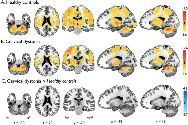
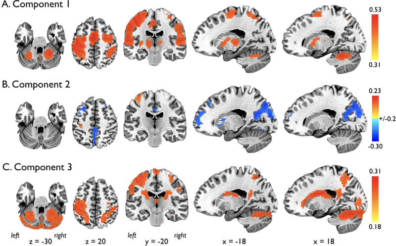
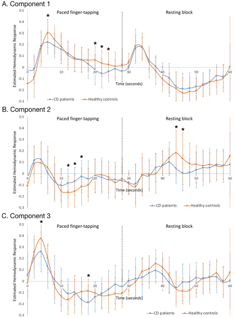
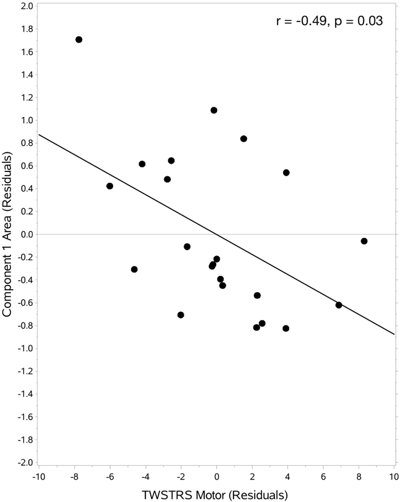
Neuroimaging studies using functional magnetic resonance imaging (fMRI), which measures brain activity by detecting the changes in blood oxygenation levels, are advancing our understanding of the pathophysiology of dystonia. Neurobiological disturbances in dystonia, however, may affect neurovascular coupling and impact the interpretability of fMRI studies. We evaluated here whether the hemodynamic response patterns during a behaviorally matched motor task are altered in isolated cervical dystonia (CD). Twenty-five CD patients and 25 healthy controls (HCs) underwent fMRI scanning during a paced finger tapping task (nondystonic task in patients). Imaging data were analyzed using a constrained principal component analysis-a statistical method that combines regression analysis and principal component analysis and enables the extraction of task-related functional networks and determination of the spatial and temporal hemodynamic response patterns associated with the task performance. Data from three patients and two controls were removed due to excessive movement. No significant differences in demographics or motor performance were observed. Three task-associated functional brain networks were identified. During task performance, reduced hemodynamic responses were seen in a sensorimotor network and in a network that included key nodes of the default mode, executive control and visual networks. During rest, reductions in hemodynamic responses were seen in the cognitive/visual network. Lower hemodynamic responses within the primary sensorimotor network in patients were correlated with the increased dystonia severity. Pathophysiological disturbances in isolated CD, such as alterations in inhibitory signaling and dopaminergic neurotransmission, may impact neurovascular coupling. Not accounting for hemodynamic response differences in fMRI studies of dystonia could lead to inaccurate results and interpretations.
Keywords: BOLD; cervical dystonia; fMRI; finger tapping; hemodynamic response.
© 2019 Wiley Periodicals, Inc.
Conflict of interest statement
Conflict of interest statement
The authors report no financial or other potential conflicts of interest.
Figures
Similar articles
-
Functional activity of the sensorimotor cortex and cerebellum relates to cervical dystonia symptoms.Hum Brain Mapp. 2017 Sep;38(9):4563-4573. doi: 10.1002/hbm.23684. Epub 2017 Jun 8. Hum Brain Mapp. 2017. PMID: 28594097 Free PMC article.
-
Task-free functional MRI in cervical dystonia reveals multi-network changes that partially normalize with botulinum toxin.PLoS One. 2013 May 1;8(5):e62877. doi: 10.1371/journal.pone.0062877. Print 2013. PLoS One. 2013. PMID: 23650536 Free PMC article.
-
Disruption in cerebellar and basal ganglia networks during a visuospatial task in cervical dystonia.Mov Disord. 2017 May;32(5):757-768. doi: 10.1002/mds.26930. Epub 2017 Feb 10. Mov Disord. 2017. PMID: 28186664
-
Laminar fMRI: What can the time domain tell us?Neuroimage. 2019 Aug 15;197:761-771. doi: 10.1016/j.neuroimage.2017.07.040. Epub 2017 Jul 20. Neuroimage. 2019. PMID: 28736308 Free PMC article. Review.
-
Submillimeter-resolution fMRI: Toward understanding local neural processing.Prog Brain Res. 2016;225:123-52. doi: 10.1016/bs.pbr.2016.03.003. Epub 2016 Apr 1. Prog Brain Res. 2016. PMID: 27130414 Review.
Cited by
-
The confound of hemodynamic response function variability in human resting-state functional MRI studies.Front Neurosci. 2023 Jul 14;17:934138. doi: 10.3389/fnins.2023.934138. eCollection 2023. Front Neurosci. 2023. PMID: 37521709 Free PMC article.
References
Publication types
MeSH terms
Grants and funding
LinkOut - more resources
Full Text Sources
