. 2020 Jan 27;59(5):1818-1827.
doi: 10.1002/anie.201913029. Epub 2019 Dec 10.

3-O-Sulfation of Heparan Sulfate Enhances Tau Interaction and Cellular Uptake

Affiliations

3-O-Sulfation of Heparan Sulfate Enhances Tau Interaction and Cellular Uptake

Jing Zhao et al. Angew Chem Int Ed Engl. .

Abstract

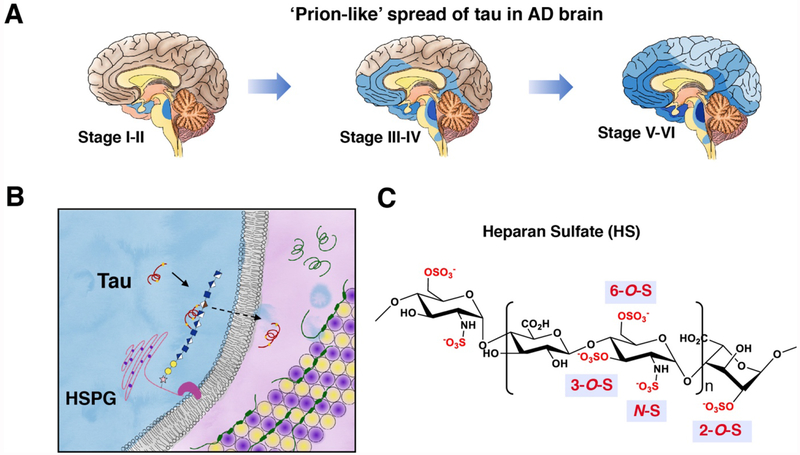
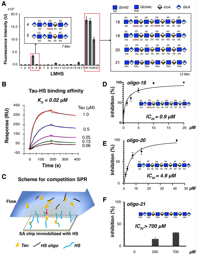
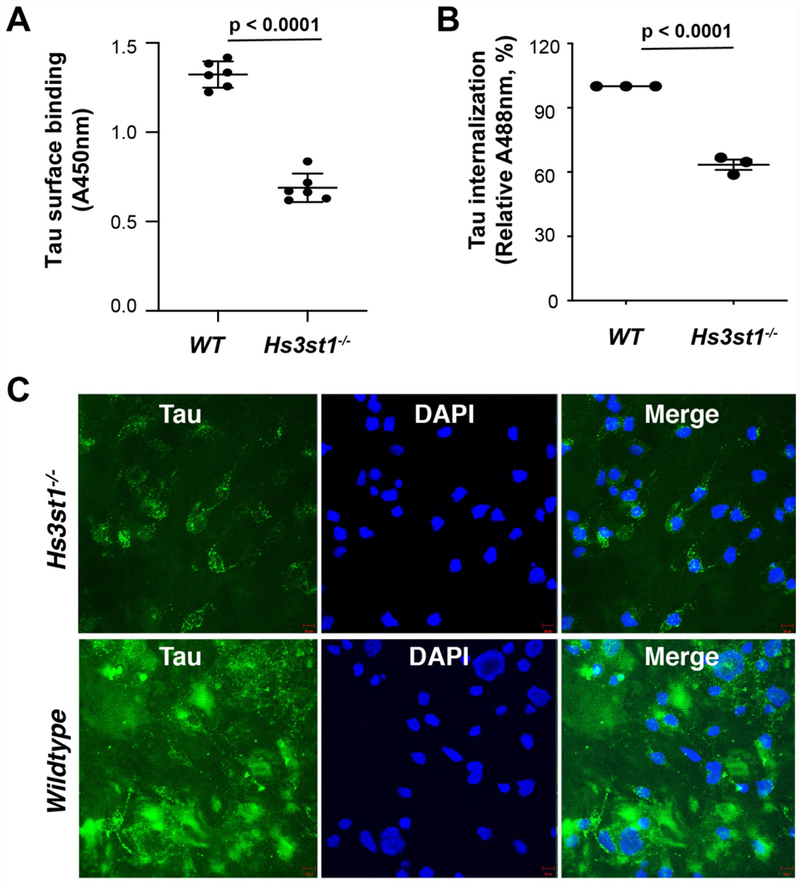
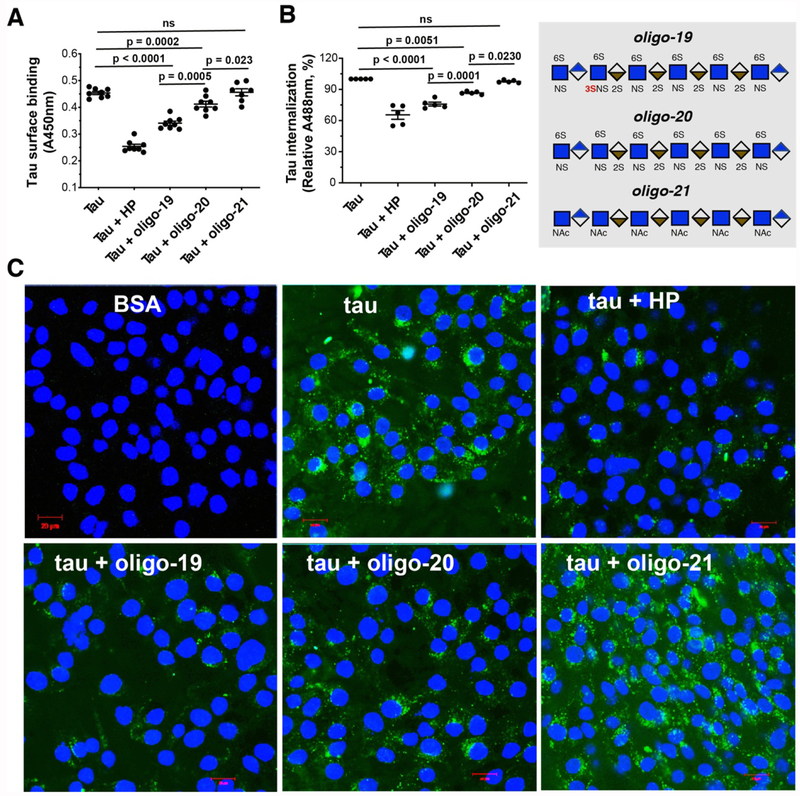
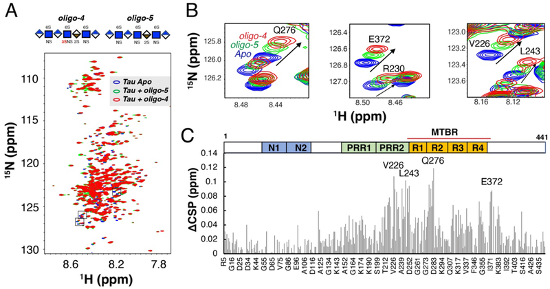
Prion-like transcellular spreading of tau in Alzheimer's Disease (AD) is mediated by tau binding to cell surface heparan sulfate (HS). However, the structural determinants for tau-HS interaction are not well understood. Microarray and SPR assays of structurally defined HS oligosaccharides show that a rare 3-O-sulfation (3-O-S) of HS significantly enhances tau binding. In Hs3st1-/- (HS 3-O-sulfotransferase-1 knockout) cells, reduced 3-O-S levels of HS diminished both cell surface binding and internalization of tau. In a cell culture, the addition of a 3-O-S HS 12-mer reduced both tau cell surface binding and cellular uptake. NMR titrations mapped 3-O-S binding sites to the microtubule binding repeat 2 (R2) and proline-rich region 2 (PRR2) of tau. Tau is only the seventh protein currently known to recognize HS 3-O-sulfation. Our work demonstrates that this rare 3-O-sulfation enhances tau-HS binding and likely the transcellular spread of tau, providing a novel target for disease-modifying treatment of AD and other tauopathies.

Keywords: Alzheimer's disease; cell surfaces; electrostatic interactions; heparan sulfate; proteins.


Figures

Figure 1.
Figure 1.. Cellular uptake of tau is mediated by HSPGs on cell surface.
(A) Prion-like spread of tau pathology (represented by blue color) in AD brain. (B) Uptake of tau mediated by the binding to heparan sulfate proteoglycans (HSPGs). Microtubules are represented by a tube composed of α- and β- tubulins (yellow and purple). (C) Primary structure and sulfation pattern of heparan sulfate.
Figure 2.
Figure 2.. Low molecular weight heparan sulfate (LMHS) array and SPR assays show the crucial role of 3-O-sulfation (3-O-S) in tau binding.
(A) 3-O-S enhances tau binding to HS in LMHS analysis. Fluorescence intensity on each spot of array was shown in a bar graph, with the monosaccharide composition/sulfation pattern drawn for the HS oligosaccharides with high fluorescence intensity (tau binding). Complete results of the LMHS array can be found in Fig. S1 and S2. (B) Binding affinity of full-length tau-HS interaction was measured to be 0.02 μM by SPR binding kinetic assay for the first time. The association and dissociation curve of different tau concentrations were fitted (black line) by a 1:1 Langmuir kinetics model in Bio-evaluation. HS from three sources (porcine brain, porcine spine and porcine intestine) were tested (Fig. S3) and only porcine intestinal HS binding is shown here. (C) Scheme for Competition SPR. (D) Oligo-19 inhibits tau-HS binding with an IC50 of 0.9 μM. (E) Oligo-20 inhibits tau-HS binding with an IC50 of 4.9 μM. (F) Oligo-21 does not inhibits tau-HS binding, with an IC50 higher than 700 μM.
Figure 3.
Figure 3.. Deletion of Hs3st1 diminishes tau cell surface binding and internalization.
(A) The Hs3st1−/− cells showed less (46.3% reduction) tau cell surface binding, compared with WT. After fixing and incubating with biotinylated full-length tau (500 ng/ml, 100 μL/well) for 90 min at RT, the cell surface bound tau was measured after incubating with Streptavidin-HRP and color development. (B) The Hs3st1−/− cells showed significantly less internalization of tau-Alexa (500 ng/ml) assessed by flow cytometry. (C) The Hs3st1−/− cells showed significantly less internalization of tau-Alexa by confocal images. The cells in 12-well plate were incubating with tau-Alexa (2 μg/ml, 500 μL/well) at 37°C for 3 h. The data shown are representative of 2-4 independent experiments.
Figure 4.
Figure 4.. 3-O-S modification enhances the inhibitory potency of HS oligo on tau-cell interaction and tau cellular uptake.
(A) HP, oligo-19 and oligo-20 inhibit tau cell surface binding by 46.3%, 28.0% and 13.0%, respectively. After fixing and incubating with biotinylated tau (500 ng/ml, 100 μL/well) without or with HP (50 ng), HS oligos (25 ng) for 90 mins at RT, the cell surface bound tau was measured after incubating with Streptavidin-HRP and color development. Oligo19 has a stronger inhibitory potency than oligo-20. Oligo-21 has no inhibition. (B) HP, oligo-19 and oligo-20 inhibit tau-Alexa (500 ng/ml) internalization assessed by flow cytometry. (C) HP, oligo-19 and oligo-20 inhibit tau internalization assessed by confocal images. The cells were incubated with tau-Alexa (2 μg/ml, 500 μL/well) without or with HP (10 μg/ml), HS oligo (2.5 μg/ml) at 37°C for 3 h. Oligo-19 has a stronger inhibitory potency than olig-20. Olig-21 has no inhibition. The data shown are representative of 2-4 independent experiments.
Figure 5.
Figure 5.. Chemical shift perturbation difference (ΔCSP) reveals specific interactions between 3-O-S and PRR2 and R2 domain of full-length tau.
(A) Overlay of 1H-15N HSQC spectra of full-length tau before (blue) and after 1:0.6 molar ratio addition of HS 7-mer oligo-5 (green) and HS 7-mer oligo-4 (red). (B) Zoomed-in NMR spectra of residues with biggest CSPs. (C) CSP differences (ΔCSP) reveals specific interaction between 3-O-S and tau PRR2 and R2 domain. Construct of tau is shown above the figure, PRR = proline-rich region, MTBR = microtubule binding region.

References

    1. Arriagada PV, Growdon JH, Hedley-Whyte ET, Hyman BT, Neurology 1992, 42, 631–631. - PubMed
    1. Wu JW, Herman M, Liu L, Simoes S, Acker CM, Figueroa H, Steinberg JI, Margittai M, Kayed R, Zurzolo C, et al., J. Biol. Chem 2013, 288, 1856–1870. - PMC - PubMed
    1. Frost B, Jacks RL, Diamond MI, J. Biol. Chem 2009, 284, 12845–12852. - PMC - PubMed
    1. Liu L, Drouet V, Wu JW, Witter MP, Small SA, Clelland C, Duff K, PLoS One 2012, 7, 1–9. - PMC - PubMed
    1. De Calignon A, Polydoro M, Suárez-Calvet M, William C, Adamowicz DH, Kopeikina KJ, Pitstick R, Sahara N, Ashe KH, Carlson GA, et al., Neuron 2012, 73, 685–697. - PMC - PubMed

Publication types