

Application of deep learning to the classification of uterine cervical squamous epithelial lesion from colposcopy images
- PMID: 31692958
- PMCID: PMC6826263
- DOI: 10.3892/mco.2019.1932
Application of deep learning to the classification of uterine cervical squamous epithelial lesion from colposcopy images
Abstract
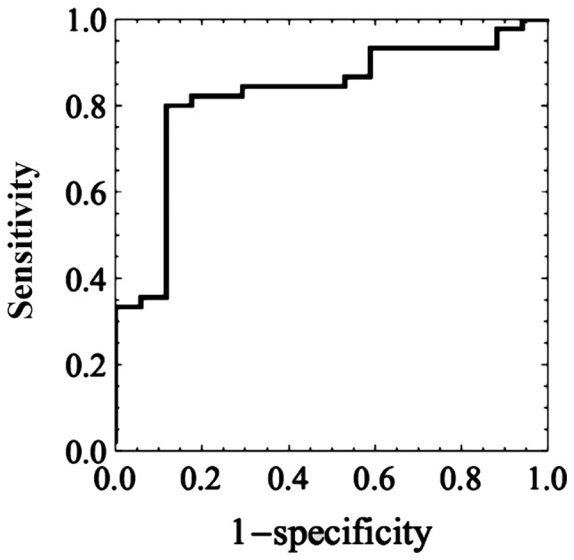
The aim of the present study was to explore the feasibility of using deep learning as artificial intelligence (AI) to classify cervical squamous epithelial lesions (SIL) from colposcopy images. A total of 330 patients who underwent colposcopy and biopsy by gynecologic oncologists were enrolled in the current study. A total of 97 patients received a pathological diagnosis of low-grade SIL (LSIL) and 213 of high-grade SIL (HSIL). An original AI-classifier with 11 layers of the convolutional neural network was developed and trained. The accuracy, sensitivity, specificity and Youden's J index of the AI-classifier and oncologists for diagnosing HSIL were 0.823 and 0.797, 0.800 and 0.831, 0.882 and 0.773, and 0.682 and 0.604, respectively. The area under the receiver-operating characteristic curve was 0.826±0.052 (mean ± standard error), and the 95% confidence interval 0.721-0.928. The optimal cut-off point was 0.692. Cohen's Kappa coefficient for AI and colposcopy was 0.437 (P<0.0005). The AI-classifier performed better than oncologists, although not significantly. Although further study is required, the clinical use of AI for the classification of HSIL/LSIL from by colposcopy may be feasible.
Keywords: artificial intelligence; cervical cancer; cervical intraepithelial neoplasia; colposcopy; deep learning.
Copyright: © Miyagi et al.
Figures
References
-
- Müller VC, Bostrom N. Springer; Berlin: 2016. Future progress in artificial intelligence: A survey of expert opinion. In: Fundamental Issues of Artificial Intelligence; pp. 555–572.
-
- Kyrgiou M, Tsoumpou I, Vrekoussis T, Martin-Hirsch P, Arbyn M, Prendiville W, Mitrou S, Koliopoulos G, Dalkalitsis N, Stamatopoulos P, Paraskevaidis E. The up-to-date evidence on colposcopy practice and treatment of cervical intraepithelial neoplasia: The Cochrane colposcopy and cervical cytopathology collaborative group (C5 group) approach. Cancer Treat Rev. 2006;32:516–523. doi: 10.1016/j.ctrv.2006.07.008. - DOI - PubMed
LinkOut - more resources
Full Text Sources