. 2019 Nov 27;141(47):18851-18861.
doi: 10.1021/jacs.9b10376. Epub 2019 Nov 18.

Excision of 5-Carboxylcytosine by Thymine DNA Glycosylase

Affiliations

Excision of 5-Carboxylcytosine by Thymine DNA Glycosylase

Lakshmi S Pidugu et al. J Am Chem Soc. .

Abstract

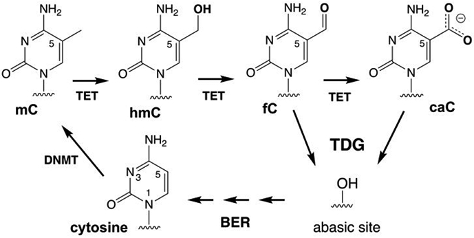
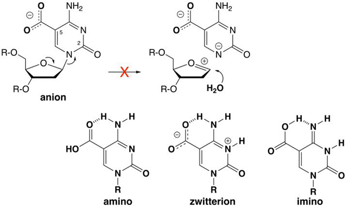
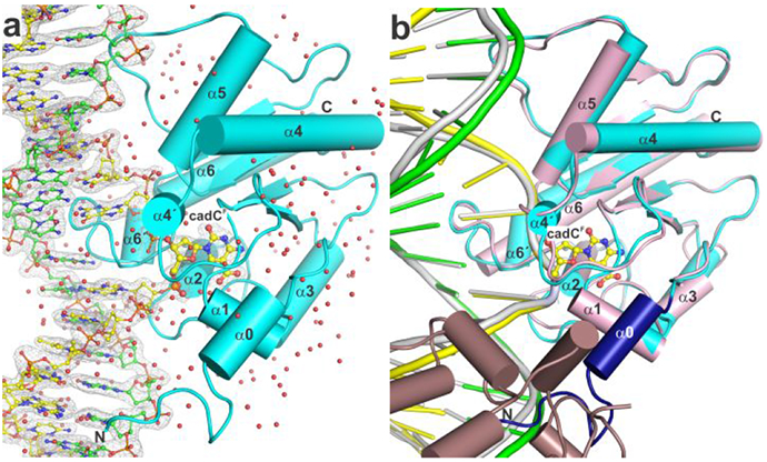
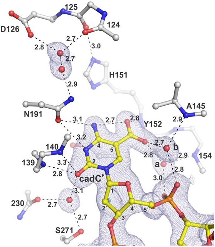
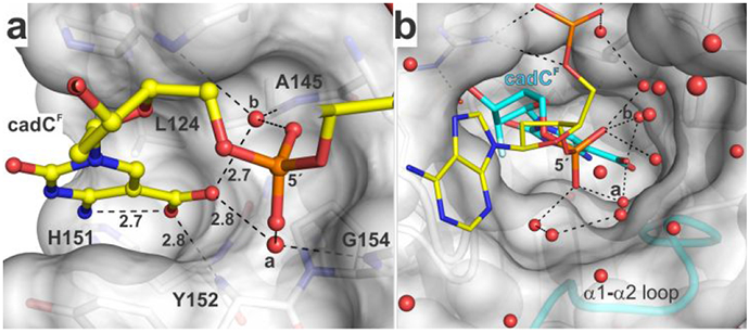
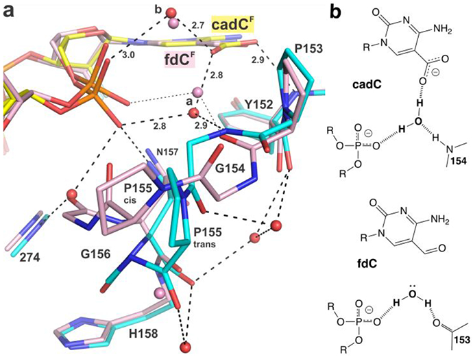
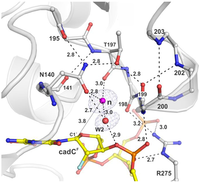
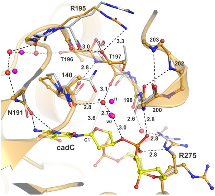
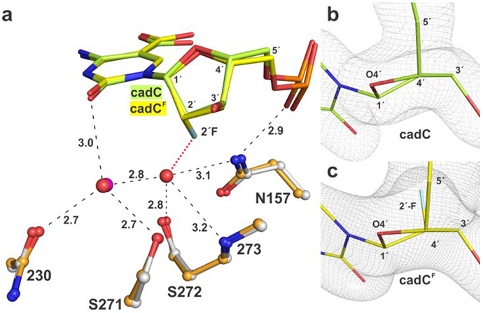
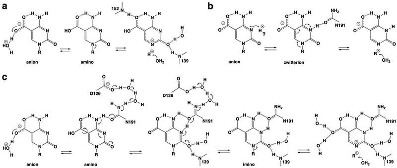
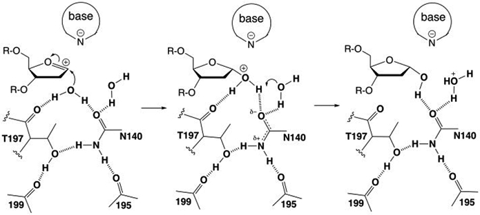
5-Methylcytosine (mC) is an epigenetic mark that is written by methyltransferases, erased through passive and active mechanisms, and impacts transcription, development, diseases including cancer, and aging. Active DNA demethylation involves TET-mediated stepwise oxidation of mC to 5-hydroxymethylcytosine, 5-formylcytosine (fC), or 5-carboxylcytosine (caC), excision of fC or caC by thymine DNA glycosylase (TDG), and subsequent base excision repair. Many elements of this essential process are poorly defined, including TDG excision of caC. To address this problem, we solved high-resolution structures of human TDG bound to DNA with cadC (5-carboxyl-2'-deoxycytidine) flipped into its active site. The structures unveil detailed enzyme-substrate interactions that mediate recognition and removal of caC, many involving water molecules. Importantly, two water molecules contact a carboxylate oxygen of caC and are poised to facilitate acid-catalyzed caC excision. Moreover, a substrate-dependent conformational change in TDG modulates the hydrogen bond interactions for one of these waters, enabling productive interaction with caC. An Asn residue (N191) that is critical for caC excision is found to contact N3 and N4 of caC, suggesting a mechanism for acid-catalyzed base excision that features an N3-protonated form of caC but would be ineffective for C, mC, or hmC. We also investigated another Asn residue (N140) that is catalytically essential and strictly conserved in the TDG-MUG enzyme family. A structure of N140A-TDG bound to cadC DNA provides the first high-resolution insight into how enzyme-substrate interactions, including water molecules, are impacted by depleting the conserved Asn, informing its role in binding and addition of the nucleophilic water molecule.


Figures

Figure 1.
Figure 1.
Methylation of cytosine and active demethylation by the TET-TDG-BER pathway in vertebrates.
Figure 2.
Figure 2.
The caC base is a monoanion at physiological pH and N-glycosyl bond hydrolysis is likely precluded by the poor leaving-group (LG) quality of a departing caC dianion. For clarity, the focus is on LG departure in a stepwise mechanism (oxacarbenium ion intermediate), without showing details for nucleophile addition (see text). Leaving group quality is improved by protonation of the caC anion to give a neutral species (amine, zwitterion, or imino), as shown by previously calculated N1 acidities, where the free energy of deprotonation (kcal mol−1) in water is 27.7 for the caC anion and 20.5, 13.3, and 16.0 for the amino, zwitterion, and imino forms of neutral caC, respectively.
Figure 3.
Figure 3.
High-resolution structure of TDG bound to G·caC DNA. (a) Crystal structure of TDG82-308 bound to DNA with cadCF in its active site (1.55 Å; PDB ID 6U17). TDG82-308 is cyan, water molecules are red spheres, the target DNA strand (with cadCF) is yellow and the complementary strand green (with N, blue; O, red; P, orange). The 2FoFc electron density map, contoured at 1.0σ, is shown for DNA. (b) Alignment of our new structure and a previous structure of TDG111-308 bound to cadCF DNA (3.01 Å; PDB ID 3UOB). Coloring for the new structure is the same except that residues 107-122 are dark blue. The previous structure exhibited 2:1 binding, one TDG subunit (pink) bound at a G·caC site and the adjacent subunit (dirty violet) at a nonspecific site.
Figure 4.
Figure 4.
Structure of TDG82-308 bound to DNA with cadCF flipped into the active site. The 2FoFc electron density map, contoured at 1.0σ, is shown for cadCF DNA and water molecules. Dashed lines represent hydrogen bonds, with interatomic distances (Å) shown. Water molecules contacting the carboxylate of cadC are labeled (a, b). The 2′-F substituent, colored cyan, is partially obscured by C2′, though its electron density is visible.
Figure 5.
Figure 5.
Binding pocket for the caC carboxylate and water molecules that contact this group and populate a channel to the surface. (a) Binding pocket for carboxyl of caC as seen in the structure of TDG82-308 bound to cadCF DNA. Select residues of TDG are shown in surface and stick format, with formatting otherwise similar to that in Figure 4. The 2′-F substituent is not shown (for clarity). (b) Water-filled channel from the TDG active site to its surface. The hydrogen bonding network (dashed lines) could facilitate transfer of a solvent proton to caC. Water molecules that contact the carboxylate of cadC are labeled (a, b).
Figure 6.
Figure 6.
Substrate dependent conformational switch. (a) The new structure of TDG82-308 (cyan) bound to cadCF DNA (yellow) superimposed with the previous structure of TDG82-308 bound to fdCF DNA (pink) (PDB ID 5T2W). Red (or pink) spheres represent water molecules. For cadC DNA, the water molecule of interest receives a hydrogen bond from a backbone nitrogen (G154), while for fdC DNA, the corresponding water (pink) donates a hydrogen bond to a backbone oxygen (P153). Cis-trans isomerization is shown for P155 (b) Effect of the conformational switch on hydrogen bond interactions of the ordered water molecule and its capacity to provide a hydrogen bond to the carboxylate of caC.
Figure 7.
Figure 7.
Coordination of the nucleophilic water molecule for TDG82-308 bound to cadCF DNA (PDB ID: 6U17). The 2FoFc electron density map, contoured at 1.0σ, is shown for water molecules only (some omitted for clarity). Dashed lines represent hydrogen bonds with interatomic distances (in Å) shown (otherwise ≤3.3 Å). The nucleophile (magenta sphere) and electrophile (C1′) are separated by 3.8 Å (thin dotted line).
Figure 8.
Figure 8.
Effects of removing the N140 carboxamide. Structure of N140A-TDG82-308 (wheat) bound to DNA (light green) with cadC flipped into its active site (PDB ID: 6U16). Aligned to this is the structure of TDG82-308 (white) bound to cadCF DNA (PDB ID: 6U17). Water molecules are red spheres for N140A-TDG82-308 and magenta for TDG82-308. The putative nucleophile for TDG82-308 is labeled “n”; a corresponding water displaced by 1 Å for N140A-TDG82-308 is labeled “*” and its distance from the electrophile (C1′) is 3.6 Å (thin dotted line). The second water that contacts N140 of TDG82-308 is labeled “W2”.
Figure 9.
Figure 9.
Effect of 2′-F substituent on the conformation of cadC flipped into the active site of N140A-TDG82-308. (a) Alignment of structures of N140A-TDG82-308 bound to DNA containing cadC (lime) or cadCF (yellow) reveals little difference in cadC conformation. Water molecules are red spheres for cadC and magenta for cadCF; one water is seen for cadC but not cadCF, due likely to steric hindrance with 2′-F (red dotted line). Dashed lines represent hydrogen bonds with interatomic distances (Å). (b, c) Close-up view of cadC and cadCF nucleotides, flipped into the N140A-TDG82-308 active site, indicate that 2′-F has a minor effect on sugar pucker (C1′-exo, slight O4′-exo). View is along a C4′-C2′ axis with C4′ in foreground. 2FoFC electron density maps are contoured at 1.0σ.
Figure 10.
Figure 10.
Potential mechanisms for acid-catalyzed excision of caC by TDG, with a focus on steps leading to N-glycosyl bond cleavage. We note that subsequent steps, including potential mechanisms for nucleophile addition and product release, are not shown. (a) Activation of the caC anion through protonation at its carboxylate to give the neutral caC amino; the mechanism involves proton shuttling through one of the two water molecules shown here to contact the caC carboxylate. (b) Activation of the caC anion through protonation at N3 to give the neutral zwitterion; the “?” indicates that the source of the requisite proton is unclear. (c) A new mechanism for activation of the caC anion involves water-mediated protonation of the carboxylate to give the neutral caC amino and its conversion to the caC imino, mediated by N191 and its interaction (through water molecules) with a general base, D126. For all three mechanisms, some interactions with water molecules or TDG groups are shown only in the most relevant steps or not at all (for clarity).
Figure 11.
Figure 11.
Potential role for the conserved Asn and Thr residues of TDG-MUG enzymes in positioning the nucleophilic water molecule, stabilizing positive charge upon nucleophile addition, and facilitating proton transfer to another water molecule. The structural interactions are shown in Figure 7. Labels denote residues in human TDG.

References

    1. Kohli RM; Zhang Y, TET enzymes, TDG and the dynamics of DNA demethylation. Nature 2013, 502 (7472), 472–9. - PMC - PubMed
    1. Drohat AC; Coey CT, Role of Base Excision "Repair" Enzymes in Erasing Epigenetic Marks from DNA. Chem Rev 2016, 116 (20), 12711–12729. - PMC - PubMed
    1. Tahiliani M; Koh KP; Shen Y; Pastor WA; Bandukwala H; Brudno Y; Agarwal S; Iyer LM; Liu DR; Aravind L; Rao A, Conversion of 5-methylcytosine to 5-hydroxymethylcytosine in mammalian DNA by MLL partner TET1. Science 2009, 324 (5929), 930–5. - PMC - PubMed
    1. He YF; Li BZ; Li Z; Liu P; Wang Y; Tang Q; Ding J; Jia Y; Chen Z; Li L; Sun Y; Li X; Dai Q; Song CX; Zhang K; He C; Xu GL, Tet-Mediated Formation of 5-Carboxylcytosine and Its Excision by TDG in Mammalian DNA. Science 2011, 333 (6047), 1303–1307. - PMC - PubMed
    1. Ito S; Shen L; Dai Q; Wu SC; Collins LB; Swenberg JA; He C; Zhang Y, Tet Proteins Can Convert 5-Methylcytosine to 5-Formylcytosine and 5-Carboxylcytosine. Science 2011, 333 (6047), 1300–1303. - PMC - PubMed

Publication types

MeSH terms