. 2019 Nov 18;58(22):15455-15465.
doi: 10.1021/acs.inorgchem.9b02530. Epub 2019 Nov 6.

Spectroscopic Evidence for Electronic Control of Heme Hydroxylation by IsdG

Affiliations

Spectroscopic Evidence for Electronic Control of Heme Hydroxylation by IsdG

Matthew A Conger et al. Inorg Chem. .

Abstract

Staphylococcus aureus IsdG catalyzes a unique trioxygenation of heme to staphylobilin, and the data presented in this article elucidate the mechanism of the novel chemical transformation. More specifically, the roles of the second-sphere Asn and Trp residues in the monooxygenation of ferric-peroxoheme have been clarified via spectroscopic characterization of the ferric-azidoheme analogue. Analysis of UV/vis absorption data quantified the strength of the hydrogen bond that exists between the Asn7 side chain and the azide moiety of ferric-azidoheme. X-band electron paramagnetic resonance data were acquired and analyzed, which revealed that this hydrogen bond weakens the π-donor strength of the azide, resulting in perturbations of the Fe 3d based orbitals. Finally, nuclear magnetic resonance characterization of 13C-enriched samples demonstrated that the Asn7···N3 hydrogen bond triggers partial porphyrin to iron electron transfer, resulting in spin density delocalization onto the heme meso carbons. These spectroscopic experiments were complemented by combined quantum mechanics/molecular mechanics computational modeling, which strongly suggested that the electronic structure changes observed for the N7A variant arose from loss of the Asn7···N3 hydrogen bond as opposed to a decrease in porphyrin ruffling. From these data a fascinating picture emerges where an Asn7···N3 hydrogen bond is communicated through four bonds, resulting in meso carbons with partial cationic radical character that are poised for hydroxylation. This chemistry is not observed in other heme proteins because Asn7 and Trp67 must work in concert to trigger the requisite electronic structure change.


Figures
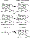
Figure 1.
Figure 1.
IsdG degrades heme to the trioxygenated tetrapyrrole product, staphylobilin, via ferric−peroxoheme and meso-hydroxyheme intermediates (top). The goal of this work is to elucidate the chemical mechanism for the ferric−peroxoheme to meso-hydroxyheme conversion. In order to achieve this goal, selectively 13C enirched heme was biosynthesized from [5-13C]-δ-aminolevulinic acid (bottom).
Figure 2.
Figure 2.
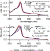
UV/vis Abs detected titrations of azide into WT IsdG−heme (top) and N7A IsdG−heme (bottom) in 50 mM Tris pH 7.4 and 150 mM NaCl. The traces represent samples with 0 mM (solid red), 200 mM (solid purple), and intermediate concentrations (dashed black) of azide. The UV/vis Abs detected titrations were fit to eq 1 yielding Kd values of 0.6 ± 0.1 and 2.2 ± 0.1 mM for WT and N7A IsdG−heme−N3, respectively. The N7A substitution decreases IsdG−heme azide affinity 3-fold.
Figure 3.
Figure 3.
Experimental (solid lines) and simulated (dashed lines) X-band (9.38 GHz) EPR spectra of WT and N7A IsdG−heme−N3 in 125 mM KPi pH 7.4 acquired with a microwave power of 63 μW, 100 kHz modulation frequency, 10 G modulation amplitude, and a time constant of 5.12 ms. Both spectra arise from at least two distinct low-spin ferric heme species (blue and violet dotted lines and brackets).
Figure 4.
Figure 4.
1H NMR spectra (25 °C) of WT (black line) and N7A (red line) IsdG−heme−N3 in 20 mM NaPi pH 7.4, 100 mM azide, and 10% D2O (v/v). 1H resonances consistent with either a 2Eg or 2B2g ground state are not observed for WT enzyme. 1H resonances consistent with a 2Eg ground state are observed for N7A IsdG−heme−N3. These data strongly suggest that the N7A substitution fundamentally changes the IsdG−heme−N3 spin density distribution.
Figure 5.
Figure 5.
1H-decoupled (dashed black lines) and 1H-coupled (solid red lines) 13C NMR spectra (25 °C) of WT (top) and N7A (bottom) IsdG−heme−N3 in 20 mM NaPi pH 7.4, 100 mM azide, and 10% D2O (v/v). The heme meso (m) and four of the eight α-pyrrole (α) carbons have been 13C enriched. Resonance assignments were made on the basis of the differences between the 1H-decoupled and 1H-coupled spectra. The average heme meso 13C resonance decreases from 47.1 ppm in WT IsdG−heme−N3 to 37.5 ppm in N7A IsdG−heme−N3, which is consistent with significantly decreased spin density delocalization onto the heme meso carbons in the N7A variant.
Figure 6.
Figure 6.
1H−13C HMQC spectra (25 °C) of WT (black) and N7A (red) IsdG−heme−N3 in 20 mM NaPi pH 7.4, 100 mM azide, and 10% D2O (v/v). For each variant, three of the four expected heme meso 1H−13C cross peaks (m) are detectable. These data provide further support for the 13C resonance assignments presented in Figure 5.
Figure 7.
Figure 7.
Structural alignment of the PBE/TZP + AMBER95 QM/MM-optimized structures of WT (light green) and N7A (light blue) IsdG−heme−CN. The PBE/TZP region included parts of the Asn7/Ala7, His77, heme, and cyanide residues. The N7A substitution does not significantly influence the magnitude of heme ruffling within the IsdG active site.
Figure 8.
Figure 8.
Spectroscopic data presented in this article strongly suggest that a hydrogen bond between Asn7 and the peroxo ligand stabilizes the Fe 3dxz and 3dyz based molecular orbitals without changing the electronic ground state (top). This iron electronic structure change alters spin density (gray dots) of the porphyrin ligand, resulting in meso carbons with electrophilic radical character (bottom).
Figure 9.
Figure 9.
Data presented in this article have revealed how Asn7 and Trp67 work in concert to promote IsdG-catalyzed heme hydroxylation (top). A steric interaction between Trp67 and the porphyrin ligand pushes the β- and δ-meso carbons toward the peroxo ligand and turns on mixing of the Fe 3dxy and porphyrin a2u orbitals (middle). A hydrogen bond between Asn7 and the peroxo ligand orients the bent peroxo ligand along the β-/δ-meso carbon axis and increases the contribution of the Fe 3dxy orbital to the singly occupied molecular orbital of the complex (bottom). The combination of these two perturbations results in spin density delocalization onto the heme meso carbons and an electronic driving force for heme hydroxylation. Similar hydroxylation chemistry is not observed in canonical heme oxygenases, peroxidases, or nitrophorins because both second-sphere interactions are required to promote the rearrangement reaction catalyzed by IsdG.

References

    1. Poulos TL Heme enzyme structure and function. Chem. Rev. 2014, 114, 3919–3962. - PMC - PubMed
    1. Matsui T; Unno M; Ikeda-Saito M Heme oxygenase reveals its strategy for catalyzing three successive oxygenation reactions. Acc. Chem. Res. 2010, 43, 240–247. - PubMed
    1. Wilks A; Ikeda-Saito M Heme utilization by pathogenic bacteria: not all pathways lead to biliverdin. Acc. Chem. Res. 2014, 47, 2291–2298. - PMC - PubMed
    1. Reniere ML; Ukpabi GN; Harry SR; Stec DF; Krull R; Wright DW; Bachmann BO; Murphy ME; Skaar EP The IsdG-family of haem oxygenases degrades haem to a novel chromophore. Mol. Microbiol. 2010, 75, 1529–1538. - PMC - PubMed
    1. Nambu S; Matsui T; Goulding CW; Takahashi S; Ikeda-Saito M A new way to degrade heme: the Mycobacterium tuberculosis enzyme MhuD catalyzes heme degradation without generating CO. J. Biol. Chem. 2013, 288, 10101–10109. - PMC - PubMed