. 2020 Jan 3;19(1):360-370.
doi: 10.1021/acs.jproteome.9b00575. Epub 2019 Dec 2.

Changes to the TDP-43 and FUS Interactomes Induced by DNA Damage

Affiliations

Changes to the TDP-43 and FUS Interactomes Induced by DNA Damage

Tetsuya Kawaguchi et al. J Proteome Res. .

Abstract

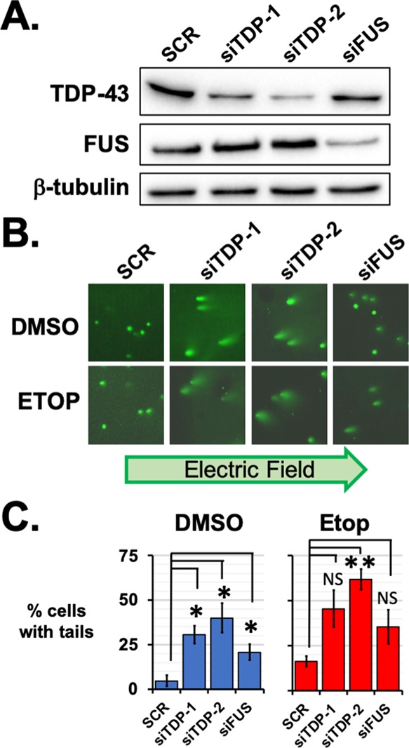
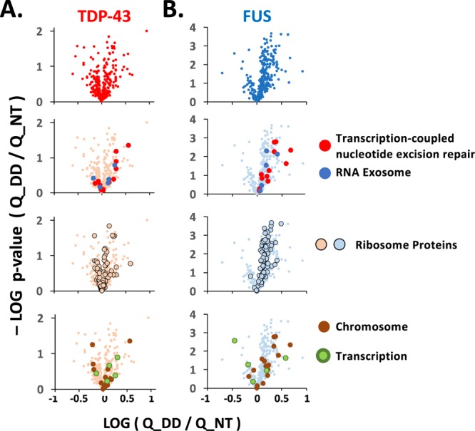
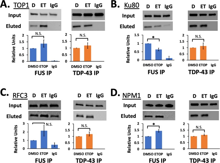
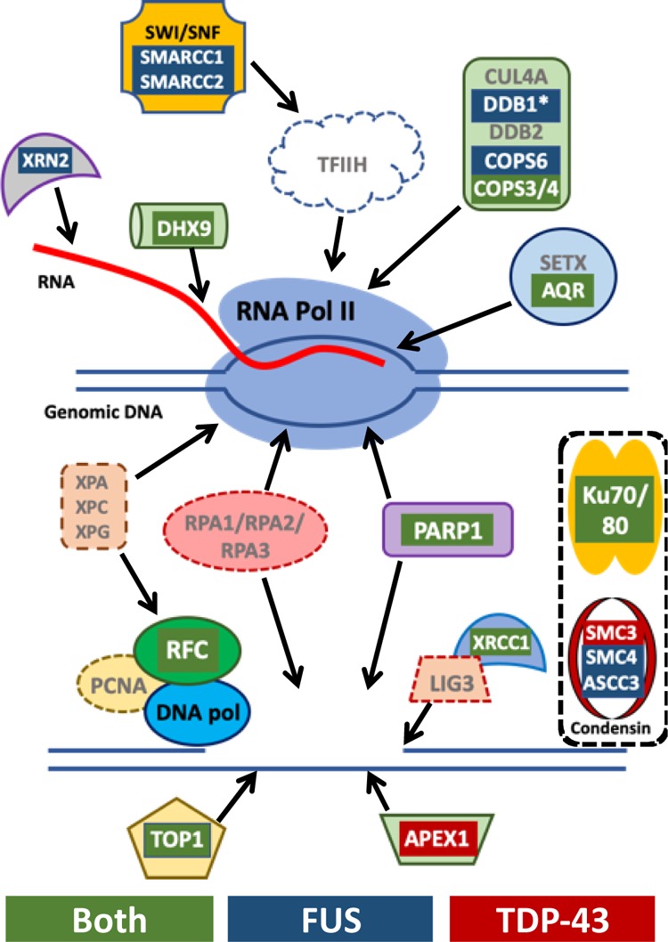
The RNA-binding proteins TDP-43 and FUS are tied as the third leading known genetic cause for amyotrophic lateral sclerosis (ALS), and TDP-43 proteopathies are found in nearly all ALS patients. Both the natural function and contribution to pathology for TDP-43 remain unclear. The intersection of functions between TDP-43 and FUS can focus attention for those natural functions mostly likely to be relevant to disease. Here, we compare the role played by TDP-43 and FUS, maintaining chromatin stability for dividing HEK293T cells. We also determine and compare the interactomes of TDP-43 and FUS, quantitating changes in those before and after DNA damage. Finally, selected interactions with known importance to DNA damage repair were validated by co-immunoprecipitation assays. This study uncovered TDP-43 and FUS binding to several factors important to DNA repair mechanisms that can be replication-dependent, -independent, or both. These results provide further evidence that TDP-43 has an important role in DNA stability and provide new ways that TDP-43 can bind to the machinery that guards DNA integrity in cells.

Keywords: DNA damage repair; FUS; TDP-43; amyotrophic lateral sclerosis; frontal temporal dementia; transcription.


Conflict of interest statement

The authors declare no competing financial interest.

Figures

Figure 1
Figure 1
TDP-43 knockdown leads to chromatin instability. (A) By western analysis, TDP-43 protein levels were found to be lower in HEK293 cells after targeted by two siRNAs: siTDP-1 and siTDP-2. Knockdown of TDP-43 did not affect the FUS expression, nor did FUS knockdown with siFUS affect TDP-43 protein levels. Knockdown results were compared with those after transfection of an siRNA with a scrambled sequence, SCR. (B) Comet tail assays were performed for SCR, siTDP-1, siTDP-2, and siFUS-treated HEK293 cells after treatment with etoposide or DMSO as a vehicle control. Comet tails comprising fragmented DNA were observed for siTDP-1 and siTDP-2 knockdowns after DMSO or 5 μM etoposide treatment for 1 h. Comet tails were also observed after etoposide treatment of cells with FUS knocked down with siFUS. (C) For each treatment, the percentage of cells with comet tails was counted from 40 to 140 cells and three biological replicates. Error bars indicate the standard error about the mean of replicates. * indicates p < 0.05 and ** indicates p < 0.005 with Student’s t-test assuming equal variances and compared to the SCR-treated samples.
Figure 2
Figure 2
Interactions of FUS and TDP-43 are affected by DNA damage. (A) AE-MS was performed on HeLa-Kyoto cells stably expressing LAP-tagged TDP-43 or FUS and treated with either DMSO or 5 μM etoposide for 1 h. After enrichment with anti-GFP camelid antibodies, interactors were digested in solution for LC–MS/MS. LFQ analysis was used to determine enrichment above GFP or LAP-SARS control proteins (p < 0.05, Student’s t-test; N = 4 for each treatment and control). (B) Stable LAP-FUS (left) or LAP-TDP43 (right) cell lines were found by western analysis to express the tagged proteins below the endogenous levels and neither the tagged nor endogenous protein levels were affected by 1 h of etoposide treatment. (C) Heat maps show changes to interactions shared by both TDP-43 and FUS (left, N = 316), FUS only (center, N = 289), or TDP-43 only (right, N = 139). Fold changes are shown as increases (green) or decreases (red). Blue bars to the right of each heat map indicate significant changes (p ≤ 0.05, Student’s t-test). The Venn diagram summarizes the number of interactors shared by TDP-43 or FUS or unique to those enrichments. (D) Complex network analysis was performed for interactors with either TDP-43 (left, red) or FUS (blue, right). The log10 of the p-value for the significance of enrichment is plotted for each GO term. A list of interactors for each GO term can be found in Table 1.
Figure 3
Figure 3
Interactions with DNA repair proteins are affected by DNA damage. Changes in enrichment determined by LFQ analysis are shown as volcano plots with the log10 of the fold change after etoposide treatment (x-axis) vs the log10 of p-values of the change for interactors shared by TDP-43 (A) and FUS (B). At the top are changes in all shared interactors detected at levels significantly above controls. Second, interactors are superimposed from the RNA exosome and transcription-coupled nucleotide repair machinery, including the RFC complex. Third, interactors that are members of the cytosolic and mitochondrial ribosomes are shown. Last are proteins classified as chromatin-bound and the few transcription-related proteins shared by FUS and TDP-43. The identity of the interactors plotted is found in Table 1. Interactors unique to TDP-43 or FUS are included in Figure 1B,C and Table 1.
Figure 4
Figure 4
TDP-43 binds DNA damage repair proteins. co-IP was performed for LAP-tagged FUS (blue) and TDP-43 (orange). Western blots show inputs and eluted proteins for TOP1 (A), Ku80 (B), RFC3 (C), and NPM1 (D). Levels of protein in western analyses were quantified and then normalized to western blots for the input samples and for the eluted LAP-FUS or LAP-TDP43. Only changes for LAP-FUS interacting with Ku80 and NPM1 were significant (p < 0.05, Student’s t-test assuming equal variances). Each western blot includes samples treated with DMSO (D), 5 μM etoposide for 1 h (ET), and pulldown with a negative control nonspecific IgG antibody (IgG). Error bars show standard error from three or four biological replicates.
Figure 5
Figure 5
Summary of TDP-43 and FUS interactors associated in transcription-coupled DNA repair. Several of the key factors in the transcription-coupled repair pathway are depicted and interactors identified by AE-MS are highlighted as red for those bound to TDP-43, blue for those bound to FUS, and green for those bound to both. Additional key factors not found in AE-MS to be enriched above controls are indicated by the gray text. The top diagram indicates factors that interact with or recruited by the stalled polymerase at sites of DNA damage. Below that are factors subsequently recruited to repair the damaged DNA. Also shown are the bound complexes Ku70/Ku80 and condensin that are known to play important roles in several DNA repair pathways but their association with transcription-coupled repair remains unclear.

References

    1. Melnick M.; Gonzales P.; Cabral J.; Allen M. A.; Dowell R. D.; Link C. D. Heat shock in C. elegans induces downstream of gene transcription and accumulation of double-stranded RNA. PLoS One 2019, 14, e0206715. 10.1371/journal.pone.0206715. - DOI - PMC - PubMed
    1. Krach F.; Batra R.; Wheeler E. C.; Vu A. Q.; Wang R.; Hutt K.; Rabin S. J.; Baughn M. W.; Libby R. T.; Diaz-Garcia S.; Stauffer J.; Pirie E.; Saberi S.; Rodriguez M.; Madrigal A. A.; Kohl Z.; Winner B.; Yeo G. W.; Ravits J. Transcriptome-pathology correlation identifies interplay between TDP-43 and the expression of its kinase CK1E in sporadic ALS. Acta Neuropathol. 2018, 136, 405–423. 10.1007/s00401-018-1870-7. - DOI - PMC - PubMed
    1. Kapeli K.; Pratt G. A.; Vu A. Q.; Hutt K. R.; Martinez F. J.; Sundararaman B.; Batra R.; Freese P.; Lambert N. J.; Huelga S. C.; Chun S. J.; Liang T. Y.; Chang J.; Donohue J. P.; Shiue L.; Zhang J.; Zhu H.; Cambi F.; Kasarskis E.; Hoon S.; Ares M. Jr.; Burge C. B.; Ravits J.; Rigo F.; Yeo G. W. Distinct and shared functions of ALS-associated proteins TDP-43, FUS and TAF15 revealed by multisystem analyses. Nat. Commun. 2016, 7, 12143. 10.1038/ncomms12143. - DOI - PMC - PubMed
    1. Anderson E. N.; Gochenaur L.; Singh A.; Grant R.; Patel K.; Watkins S.; Wu J. Y.; Pandey U. B. Traumatic injury induces stress granule formation and enhances motor dysfunctions in ALS/FTD models. Hum. Mol. Genet. 2018, 27, 1366–1381. 10.1093/hmg/ddy047. - DOI - PMC - PubMed
    1. Purice M. D.; Taylor J. P. Linking hnRNP Function to ALS and FTD Pathology. Front. Neurosci. 2018, 12, 326. 10.3389/fnins.2018.00326. - DOI - PMC - PubMed

Publication types