Review
. 2020 Jan 1;318(1):L149-L164.
doi: 10.1152/ajplung.00329.2019. Epub 2019 Nov 6.

Mitochondria: at the crossroads of regulating lung epithelial cell function in chronic obstructive pulmonary disease

Affiliations
Review

Mitochondria: at the crossroads of regulating lung epithelial cell function in chronic obstructive pulmonary disease

Mahyar Aghapour et al. Am J Physiol Lung Cell Mol Physiol. .

Abstract

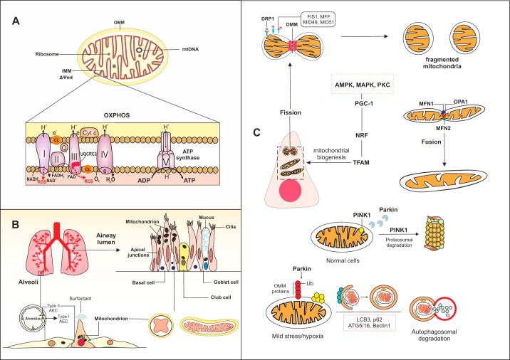
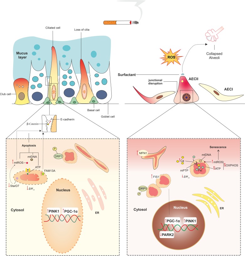
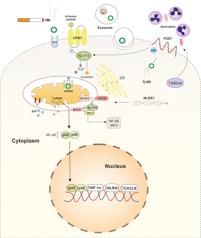
Disturbances in mitochondrial structure and function in lung epithelial cells have been implicated in the pathogenesis of various lung diseases, including chronic obstructive pulmonary disease (COPD). Such disturbances affect not only cellular energy metabolism but also alter a range of indispensable cellular homeostatic functions in which mitochondria are known to be involved. These range from cellular differentiation, cell death pathways, and cellular remodeling to physical barrier function and innate immunity, all of which are known to be impacted by exposure to cigarette smoke and have been linked to COPD pathogenesis. Next to their well-established role as the first physical frontline against external insults, lung epithelial cells are immunologically active. Malfunctioning epithelial cells with defective mitochondria are unable to maintain homeostasis and respond adequately to further stress or injury, which may ultimately shape the phenotype of lung diseases. In this review, we provide a comprehensive overview of the impact of cigarette smoke on the development of mitochondrial dysfunction in the lung epithelium and highlight the consequences for cell function, innate immune responses, epithelial remodeling, and epithelial barrier function in COPD. We also discuss the applicability and potential therapeutic value of recently proposed strategies for the restoration of mitochondrial function in the treatment of COPD.

Keywords: COPD; cigarette smoke; lung epithelial cells; mitochondrial dysfunction.


Conflict of interest statement

No conflicts of interest, financial or otherwise, are declared by the authors.

Figures

Fig. 1.
Fig. 1.
Schematic of mitochondrial function and localization in various lung epithelial cells during homeostasis. A: within the mitochondria, the double membrane with a folded inner membrane (IMM) forms cristae. The mitochondrial matrix contains circular mitochondrial DNA (mtDNA). Oxidative phosphorylation (OXPHOS), which provides the ATP required for cellular functions, occurs in this hyper-folded IMM. During OXPHOS, a proton gradient is established over the IMM through the flow of electrons through chains of enzymatic complexes (I–V), which leads to the production of reactive oxygen species (ROS), H2O, and as ATP. Cytochrome c and coenzyme Q facilitate the transfer of electrons during OXPHOS. Complexes I and III are the major sites of ROS production in the electron transport chain (ETC). Ubiquinol-cytochrome c reductase core II (UQCRC2) is a damage-sensitive protein that contributes to ROS generation in the complex III of ETC. Oxidation of cardiolipin (CL), the mitochondrial-specific lipid in the IMM, can induce oxidative damage. B: in the upper respiratory tract, the airway epithelium constitutes the first physical defensive barrier against inhaled pathogens and toxic gases and particles, whereas the type I and type II alveolar epithelial cells (AEC) that cover the alveolar surface facilitate gas exchange. C: the mitochondrial network is highly dynamic in lung epithelial cells. Various processes contribute to generation and degradation of mitochondria in the cells. These processes include fusion, fission, mitochondrial-specific autophagy (mitophagy), and mitochondrial biogenesis. During fission, recognition of activated dynamin-related protein 1 (DRP1) by surface receptors (FIS1, MFF, MID49, and MID51) leads to fragmentation of the mitochondrial outer membrane (OMM) and formation of new mitochondria. Mitochondria can form an elongated shape by fusion, in which mitochondrial membranes tether through fusion proteins in the IMM such as optic atrophy 1 (OPA1) and outer membranes mitofusin 1 (MFN1) and MFN2. Mitophagy is regulated by several proteins, including PTEN-induced kinase 1 (PINK1) and Parkin. In homeostatic conditions, translocation of PINK1 to the IMM triggers recruitment of E3 ligase Parkin, which leads to proteasomal degradation of PINK1. Upon mild stress mitochondria can undergo complete degradation by mitophagy, in which accumulation of PINK-1 in the OMM and subsequent ubiquitination of several proteins at the OMM by Parkin lead to engulfment of the damaged mitochondrion by autophagy compartments. Autophagy proteins LCB3, p62, ATG5/16, and beclin 1 facilitate the generation of phagophore and autophagosomes. Formation of new mitochondria can be triggered by activation of peroxisome proliferator-activated receptor-γ coactivator1α (PGC-1α) and downstream transcription factors, nuclear respiratory factors 1 and 2 (NRF1/2), and mitochondrial transcription factor A (TFAM). Activation of several proteins, including AMP-activated protein kinase (AMPK), mitogen-activated protein kinase (MAPK), and protein kinase C (PKC), may activate the PGC-1α signaling pathway and in turn lead to synthesis of mitochondria.
Fig. 2.
Fig. 2.
Cigarette smoke (CS) induces abnormalities in mitochondrial morphology and function in lung epithelial cells. CS exposure of lung epithelial cells results in swollen and branched mitochondria with a condensed matrix. These morphological features are accompanied by functional alterations in mitochondria, including altered oxidative phosphorylation (OXPHOS) with higher reactive oxygen species (ROS) production and lower ATP generation, depolarization of mitochondrial membrane, and impaired mitophagy, all of which cause subsequent changes at the cellular level. These changes include loss of ciliary function, increase in mucus production in the airway epithelium, and impaired production of surfactant in type II alveolar epithelial cells (AECII), leading to alveolar collapse. CS increases the permeability of the mitochondrial membranes and opens ion channels such as mitochondrial permeability transition pore (mPTP) in the inner membrane, leading to overload of iron in mitochondria and cytoplasmic accumulation of mitochondrial danger-associated molecular patterns (mtDAMPs), including Ca2+, ATP, mitochondrial ROS (mtROS), mtDNA, cardiolipin, and N-formylated peptides, further inducing oxidative damage and cell death. CS-induced mitochondrial dysfunction may contribute to a leaky manifestation of the airway epithelium by increased mtROS and subsequent weakening cellular junctions. Furthermore, increase in FAM13A has been associated with disrupted airway epithelial barrier by increasing mtROS. Excessive mtROS has also consequences for cell fate and growth, inducing permanent damage to mtDNA, leading to increased mitophagy and cell apoptosis, and irreversible arrest in cell growth. Short-term CS exposure may enhance mitochondrial biogenesis via increase in PPAR-γ coactivator1α (PGC-1α) transcript levels, whereas longer exposure times suppress this process. CS induces an imbalance in fusion/fission process by more trends to fission, leading to fragmentation of mitochondria in lung epithelial cells. CS enhances clearance of fragmented mitochondria by mitophagy through increase in PTEN-induced kinase 1 (PINK1) mRNA and protein levels in both airway epithelial cells and AECII.
Fig. 3.
Fig. 3.
Links between cigarette smoke (CS)-induced mitochondrial dysfunction and altered innate immune responses in chronic obstructive pulmonary disease (COPD). Damaged mitochondria as a result of CS exposure release their contents into to the cytoplasm, acting as damage-associated molecular patterns (DAMPs) and subsequently activating the innate immune system. Cytoplasmic levels of mitochondrial danger-associated molecular patterns (mtDAMPs), including mitochondrial reactive oxygen species (mtROS), mtDNA, ATP, cardiolipin, and Ca2+, are increased in lung epithelial cells in COPD. Increased levels of cytoplasmic mtDNA, mtROS, Ca2+, and cardiolipin activate innate immune responses by stimulation of intracellular pattern recognition receptors (PRRs), in particular the NLRP3 inflammasome. mtDNA acts as a ligand for Toll-like receptor 9 (TLR9), further activating NLRP3. mtDNA can be transferred to the neighboring cells via exosomes. N-formyl peptides encoded by mtDNA are released upon cellular damage, acting as DAMPs by activating formyl peptide receptor 1 (FPR1), which induces attraction of neutrophils to the site of damage. Extracellular ATP also activates NLRP3 via purinergic receptors P2X7 and P2Y2R. Activated NLRP3 translocates to mitochondria, which subsequently induces more damage by enhancing mtROS production. This increase in mtROS levels directly induces proinflammatory responses by increasing nuclear translocation of p65 and activating hypoxia-inducible factor-1α (HIF-1α) transcription factor. Furthermore, mtDAMPs elicit proinflammatory responses by inducing strong CXCL8 responses in airway epithelial cells, recruiting neutrophils to the site of damage. Another PRR, NLRX1, is also localized into mitochondria and interacts with mitochondrial antiviral signaling (MAVS), exerting anti-inflammatory responses by precluding interaction of NLRX1 with NLRP3 and retinoic acid-inducible gene-I (RIG-I) and subsequent activation of NF-κB and interferon regulatory transcription factor 3 (IRF3). CS reduces the expression of NLRX1 in the lung at both gene and posttranscriptional levels, perpetuating inflammation.

References

    1. Agarwal AR, Yin F, Cadenas E. Short-term cigarette smoke exposure leads to metabolic alterations in lung alveolar cells. Am J Respir Cell Mol Biol 51: 284–293, 2014. doi:10.1165/rcmb.2013-0523OC. - DOI - PubMed
    1. Agarwal AR, Zhao L, Sancheti H, Sundar IK, Rahman I, Cadenas E. Short-term cigarette smoke exposure induces reversible changes in energy metabolism and cellular redox status independent of inflammatory responses in mouse lungs. Am J Physiol Lung Cell Mol Physiol 303: L889–L898, 2012. doi:10.1152/ajplung.00219.2012. - DOI - PubMed
    1. Aghapour M, Raee P, Moghaddam SJ, Hiemstra PS, Heijink IH. Airway epithelial barrier dysfunction in chronic obstructive pulmonary disease: role of cigarette smoke exposure. Am J Respir Cell Mol Biol 58: 157–169, 2018. doi:10.1165/rcmb.2017-0200TR. - DOI - PubMed
    1. Aguilera-Aguirre L, Bacsi A, Saavedra-Molina A, Kurosky A, Sur S, Boldogh I. Mitochondrial dysfunction increases allergic airway inflammation. J Immunol 183: 5379–5387, 2009. doi:10.4049/jimmunol.0900228. - DOI - PMC - PubMed
    1. Ahmad T, Sundar IK, Lerner CA, Gerloff J, Tormos AM, Yao H, Rahman I. Impaired mitophagy leads to cigarette smoke stress-induced cellular senescence: implications for chronic obstructive pulmonary disease. FASEB J 29: 2912–2929, 2015. doi:10.1096/fj.14-268276. - DOI - PMC - PubMed

Publication types

MeSH terms