. 2020 Jan 1;123(1):70-89.
doi: 10.1152/jn.00018.2019. Epub 2019 Nov 6.

Adaptation to slope in locomotor-trained spinal cats with intact and self-reinnervated lateral gastrocnemius and soleus muscles

Affiliations

Adaptation to slope in locomotor-trained spinal cats with intact and self-reinnervated lateral gastrocnemius and soleus muscles

Dwight Higgin et al. J Neurophysiol. .

Abstract

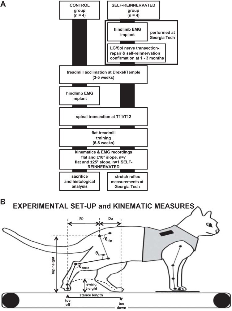
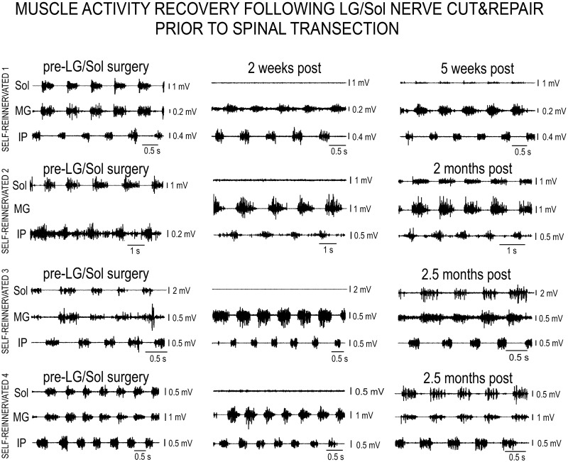
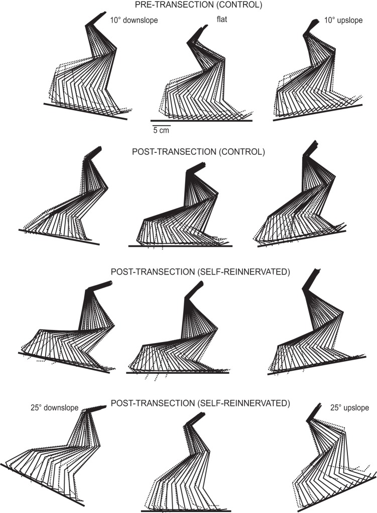
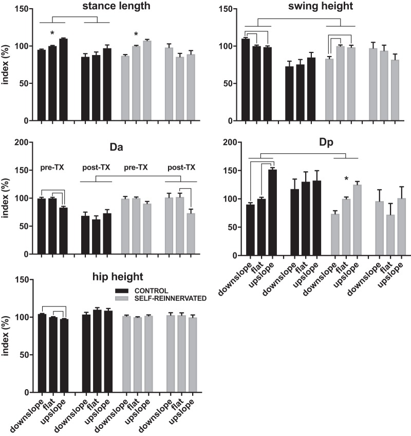
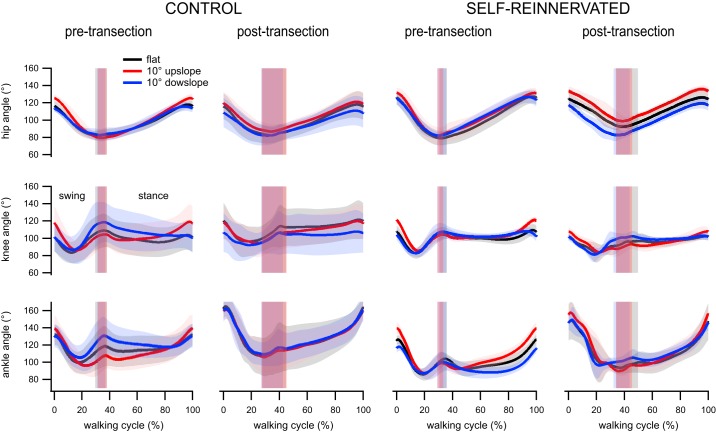
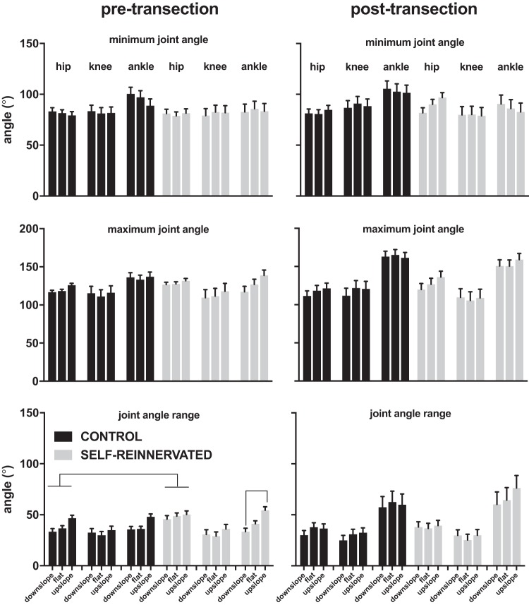
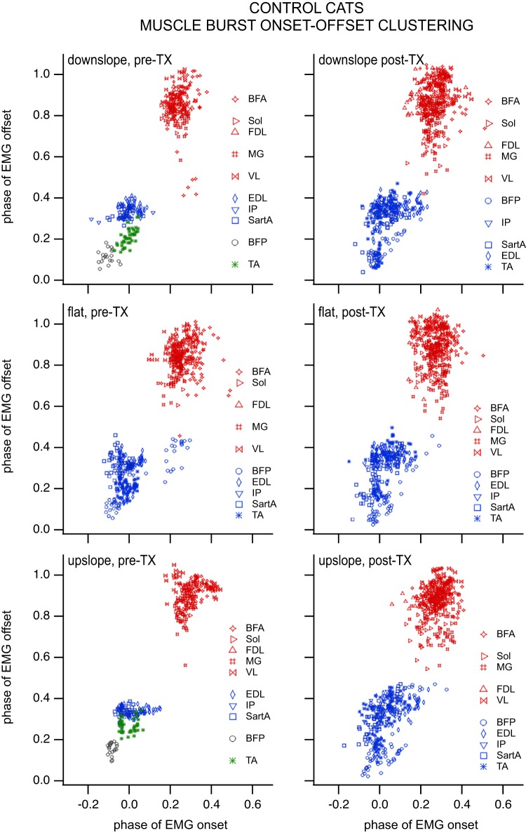
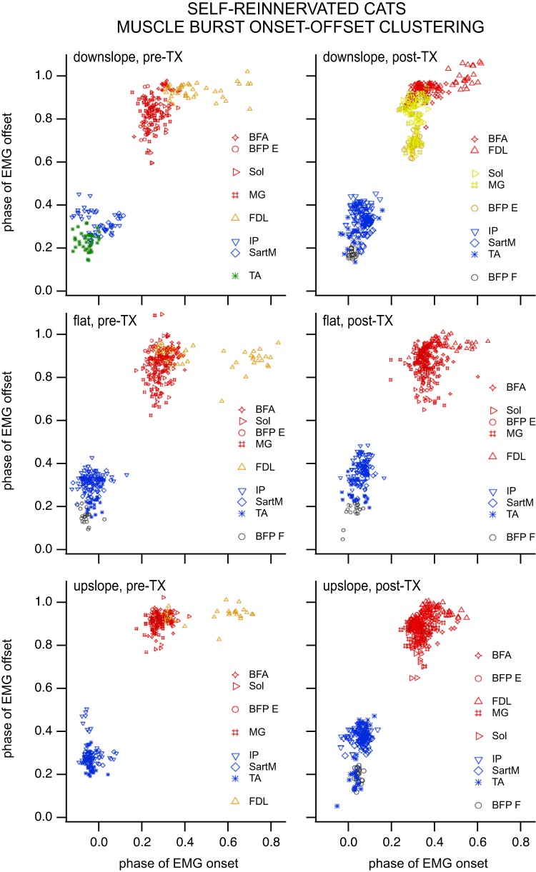
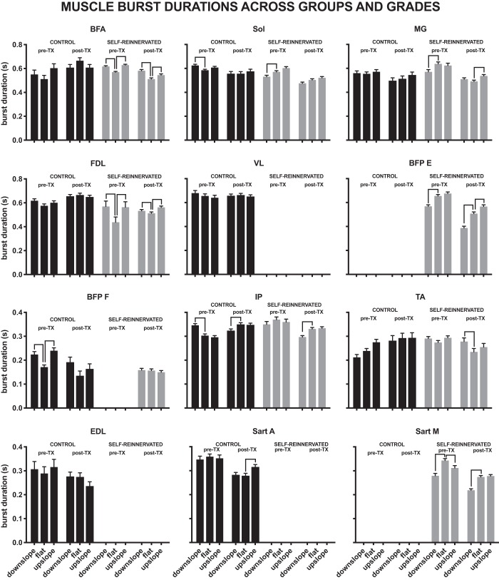
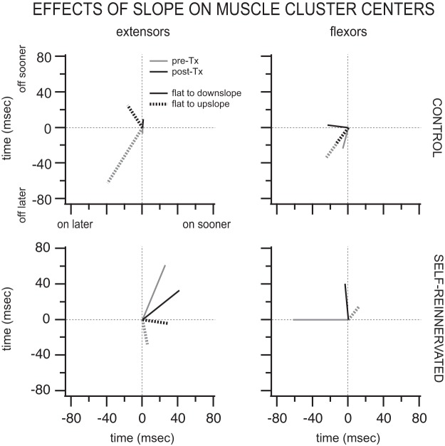
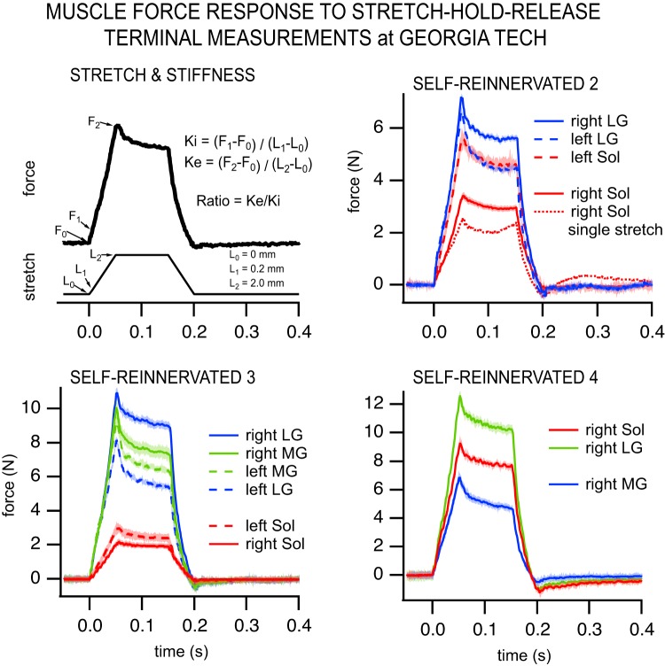
Sensorimotor training providing motion-dependent somatosensory feedback to spinal locomotor networks restores treadmill weight-bearing stepping on flat surfaces in spinal cats. In this study, we examined if locomotor ability on flat surfaces transfers to sloped surfaces and the contribution of length-dependent sensory feedback from lateral gastrocnemius (LG) and soleus (Sol) to locomotor recovery after spinal transection and locomotor training. We compared kinematics and muscle activity at different slopes (±10° and ±25°) in spinalized cats (n = 8) trained to walk on a flat treadmill. Half of those animals had their right hindlimb LG/Sol nerve cut and reattached before spinal transection and locomotor training, a procedure called muscle self-reinnervation that leads to elimination of autogenic monosynaptic length feedback in spinally intact animals. All spinal animals trained on a flat surface were able to walk on slopes with minimal differences in walking kinematics and muscle activity between animals with/without LG/Sol self-reinnervation. We found minimal changes in kinematics and muscle activity at lower slopes (±10°), indicating that walking patterns obtained on flat surfaces are robust enough to accommodate low slopes. Contrary to results in spinal intact animals, force responses to muscle stretch largely returned in both SELF-REINNERVATED muscles for the trained spinalized animals. Overall, our results indicate that the locomotor patterns acquired with training on a level surface transfer to walking on low slopes and that spinalization may allow the recovery of autogenic monosynaptic length feedback following muscle self-reinnervation.NEW & NOTEWORTHY Spinal locomotor networks locomotor trained on a flat surface can adapt the locomotor output to slope walking, up to ±25° of slope, even with total absence of supraspinal CONTROL. Autogenic length feedback (stretch reflex) shows signs of recovery in spinalized animals, contrary to results in spinally intact animals.

Keywords: afferent feedback; locomotion; reinnervation; spinal cord injury.


Conflict of interest statement

No conflicts of interest, financial or otherwise, are declared by the authors.

Figures

Fig. 1.
Fig. 1.
A: experimental timeline for the animals in the study. CONTROL animals were acclimated to walking on a flat treadmill, and baseline kinematics recordings were taken before and after muscle electromyogram (EMG) electrode implantation during walking on level and 10° upslope/downslope treadmill surfaces. Animals received from Georgia Tech (SELF-REINNERVATED) had already undergone EMG electrode implantation and lateral gastrocnemius/soleus (LG/Sol) nerve transection-repair surgery and had been evaluated to confirm muscle self-reinnervation 5 wk to 3 mo after surgery during walking on flat and ±27° (50%) grade as described in previous studies (Gregor et al. 2018; Pantall et al. 2016). Recordings were taken at Georgia Tech before and after EMG implantation and at various time points following LG/Sol nerve transection-repair. After baseline recordings were taken on all animals at Drexel/Temple, a complete spinal transection was performed at T11/T12. Animals were locomotor trained following transection for ~6–8 wk, at which point weight-bearing stepping had returned and performance plateaued. Posttransection recordings were then taken on a flat, 10° upslope, and 10° downslope grade over multiple sessions. One animal with LG/Sol self-reinnervation was also evaluated at ±25° of slope. SELF-REINNERVATED animals were then returned to Georgia Tech for terminal measurements (Lyle et al. 2016) that evaluated the stretch reflex response in the Sol and LG muscles. B: schematic of experimental apparatus showing the marker locations and kinematics measures used to evaluate the changes in locomotion. The animal’s forelimbs were standing on a fixed platform while the hindlimbs walked over the treadmill belt. Kinematics measures extracted included stance length (horizontal distance from toe down to toe off), swing height (maximum height of the metatarsophalangeal marker above the treadmill during swing), hip height (average height of the hip marker over the step), Dp (horizontal displacement of the toe marker relative to the hip at toe off), Da (horizontal displacement of the toe marker relative to the hip at toe down), and hip, knee, and ankle joint angles (θ). EMGs from several right hindlimb muscles were simultaneously acquired with the kinematics data.
Fig. 2.
Fig. 2.
Electromyographic (EMG) activity of selected muscles for Self-reinnervated animals (SELF-REINNERVATED 1–4) before lateral gastrocnemius/soleus (pre-LG/Sol) nerve cut and repair (left column), 2 wk post nerve repair (middle column), and 5 wk to 2.5 mo post nerve repair (right column). Motor activity during walking is present in the Sol muscle before nerve cut, is absent at 2 wk post-repair, and returns by ~5 wk, with the activity getting more intense by 2.5 mo. The progression shows the denervation brought on by the nerve cut and subsequent motor reinnervation.
Fig. 3.
Fig. 3.
Stick figure representations of exemplar steps at the different slopes. The stick figures represent hindlimb segments over the time course of a stride for a walking speed of 0.4 m/s. Stance phase segments are indicated by solid lines, and swing phase segments by dotted lines. First row shows 1 step at 10° downslope, flat, and 10° upslope for a CONTROL cat pretransection. Second row shows the same steps for the same animal posttransection and following locomotor training. Third row shows the same steps for a SELF-REINNERVATED animal posttransection and posttraining. Fourth row shows steps at ±25° of slope and flat for another SELF-REINNERVATED animal posttransection and posttraining. Spinal animals locomotor trained on a flat surface were able to locomote on inclined surfaces, up to ±25° of grade.
Fig. 4.
Fig. 4.
Estimated marginal means ± 95% confidence intervals of the normalized stance length, swing height, anterior displacement (Da), posterior displacement (Dp), and hip height for all animals evaluated on flat and ±10° slope of walking. Full factorial linear mixed models of each of the indexes in the pre- and posttransection conditions with group and slope as factors found significant differences (no overlap in 95% confidence interval of the estimated marginal means) in swing height and Dp between the CONTROL and SELF-REINNERVATED animals pretransection, and in Da between the same two groups posttransection. Overall, the differences between CONTROL and SELF-REINNERVATED animals were small, confirming prior results in spinally intact animals showing that lateral gastrocnemius/soleus self-reinnervation has limited effects on the kinematics of locomotion even at much higher (27°) slopes (Gregor et al. 2018). Slope had a significant effect on stance length, swing height, and Dp in spinally intact animals, but not for the spinalized animals, suggesting that the adaptation to slope may be supraspinal in origin. Significant lowering of the anterior displacement between flat and upslope was the only change with grade found in spinalized animals. *Estimated marginal means do not overlap at any of the grades; lines between groups/slopes indicate no overlap in estimated marginal means between groups/slopes. Number of steps: 20 steps for each condition/slope/subject; number of subjects: 4 CONTROL at all slopes postspinalization and flat prespinalization, 3 CONTROL at ±10° slopes prespinalization, 3 SELF-REINNERVATED at all slopes postspinalization and flat prespinalization, and 2 SELF-REINNERVATED at ±10° slope prespinalization.
Fig. 5.
Fig. 5.
Average hip, knee, and ankle angles over the walking cycle from swing onset (0%) to stance offset (100%) for animals with/without lateral gastrocnemius/soleus self-reinnervation walking on flat and ±10° slope pretransection and following spinal transection and locomotor training on a flat treadmill. Lines represent averages and the thicknesses of shading around each line indicates the 95% confidence interval. Vertical shaded bars indicate stance onset ± 95% confidence interval. The joint angles did not vary much with grades in both groups, pre- and postspinalization. The results suggest that the joint angles during flat walking will result in weight-bearing stepping at slopes of ±10°. Number of steps: 20 steps for each condition/slope/subject; number of subjects: 4 CONTROL at all slopes postspinalization and flat prespinalization, 3 CONTROL at ±10° slopes prespinalization, 3 SELF-REINNERVATED at all slopes postspinalization and flat prespinalization, and 2 SELF-REINNERVATED at ±10° slope prespinalization.
Fig. 6.
Fig. 6.
Estimated marginal means ± 95% confidence intervals of the averaged (over 20 steps) hip, knee, and ankle angle minima, maxima, and ranges during walking on flat, 10° upslope, or 10° downslope for all animals evaluated at those grades. Full factorial linear mixed models of each of the angle parameters in the pre- (left column) and posttransection (right column) conditions with group and slope as factors indicated a significant difference between the CONTROL and SELF-REINNERVATED groups for the hip range of motion pretransection. No other significant differences between groups were found. The only effect of slope was on the ankle range of motion, which was larger for upslope walking (compared with downslope) in spinally intact SELF-REINNERVATED animals (no overlap in 95% confidence interval of the estimated marginal means). Overall, the results indicate that the angular parameters were not affected by slope in either condition (intact or spinal) for animals with/without lateral gastrocnemius/soleus self-reinnervation surgery. Lines between groups/slopes indicate no overlap in estimated marginal means between groups/slopes. Number of steps: 20 steps for each condition/slope/subject; number of subjects: 4 CONTROL at all slopes postspinalization and flat prespinalization, 3 CONTROL at ±10° slopes prespinalization, 3 SELF-REINNERVATED at all slopes postspinalization and flat prespinalization, and 2 SELF-REINNERVATED at ±10° slope prespinalization.
Fig. 7.
Fig. 7.
Clustering of muscle burst onset/offset for CONTROL cats during locomotion on a flat, 10° upslope, and 10° downslope treadmill in the intact (left column) and spinalized (right column) conditions (i.e., pre- and posttransection). Each symbol represents a muscle burst onset/offset combination during a cycle. Different symbol colors indicate distinct clusters of muscle electromyographic (EMG) burst onsets/offsets based on the “graphminsspantree” procedure, whereas blank lines between muscles/symbols of the same colors in the key indicate that the main cluster identified with the graphminsspantree procedure subdivided into smaller clusters using the validity index. Biceps femoris posterior (BFP) and tibialis anterior (TA) formed distinct clusters for upslope and downslope walking pretransection but grouped with the other flexors posttransection. The number of clusters (especially those identified with the graphminsspantree procedure) went down in the spinal condition, suggesting that the spinal control of locomotion may be more primitive and produce coarser flexor and extensor groups. Number of subjects: 4 at all slopes postspinalization and flat prespinalization, 3 at ±10° slopes prespinalization. Number of bursts prespinalization (pre-TX): n = 20 for iliopsoas (IP) at each slope and BFP at ±10° slopes, n = 40 for all other muscles at ±10° slopes, whereas n = 60 of those muscles for flat, except for BFP and flexor digitorum longus (FDL), where n = 40. Number of bursts postspinalization (post-TX): n = 20 for IP, whereas n = 60 for BFP, sartorius medialis (SartA), and FDL at each slope; n = 80 for the remaining muscles at each slope (BFA, biceps femoris anterior; EDL, extensor digitorum longus; MG, gastrocnemius medialis; Sol, soleus; VL, vastus lateralis).
Fig. 8.
Fig. 8.
Clustering of muscle burst onset/offset for SELF-REINNERVATED cats during locomotion on a flat, 10° upslope, and 10° downslope treadmill in the intact (left column) and spinalized (right column) conditions (i.e., pre- and posttransection). Each symbol represents a muscle burst onset/offset combination during a cycle. Different symbol colors indicate distinct clusters of muscle electromyographic (EMG) burst onsets/offsets based on the “graphminsspantree” procedure, whereas blank lines between muscles/symbols of the same colors in the key indicate that the main cluster identified with the graphminsspantree procedure subdivided into smaller clusters using the validity index. A reduction in the number of clusters posttransection was also observed for this group. As in the CONTROL group, the biceps femoris posterior flexor burst (BFP F) and tibialis anterior (TA) tended to form distinct clusters. Interestingly, the soleus (Sol) and gastrocnemius medialis (MG) muscles formed a very distinct cluster (graphminsspantree procedure) for downslope walking posttransection. This phenomenon was not observed in animals with intact lateral gastrocnemius (LG)/Sol nerve, and prior studies have shown that the greatest deficits during slope walking in animals with LG/Sol self-reinnervation occur on downslope. The distinct clustering of both ankle extensors may be related to the loss of heteronymous stretch reflex from the LG and Sol muscles in this group, since homonymous stretch reflex recovered (Fig. 11). Number of subjects: 3 at all slopes postspinalization and flat prespinalization, 2 at ±10° slope prespinalization. Number of bursts prespinalization (pre-TX): n = 20 for BFP at each slope and sartorius medialis (SartM) at ±10° slopes, n = 40 for all other muscles at ±10° slopes, and n = 60 at flat, except for TA, which remains at n = 40 for flat. Number of bursts postspinalization (post-TX): n = 20 for BFP at each slope, n = 20 for SartM and TA, and n = 60 for the remaining muscles at each slope (BFP E, biceps femoris posterior extensor burst; EDL, extensor digitorum longus; FDL, flexor digitorum longus; IP, iliopsoas; VL, vastus lateralis).
Fig. 9.
Fig. 9.
Individual muscle burst durations for both groups (CONTROL and SELF-REINNERVATED), before (pre-TX) and after spinalization (post-TX), at each slope of walking (flat, 10° upslope, and 10° downslope). Significant differences due to slope are indicated by line segments between the bars representing the burst durations at each slope. Burst durations were not affected by a ±10° change in elevation for most of the muscles studied in animals with either an intact or lateral gastrocnemius/soleus self-reinnervation, before or after spinalization. Bars indicate the estimated marginal means ± 95% confidence interval of burst durations. Number of subjects and bursts for each bar are reported in Figs. 7 and 8.
Fig. 10.
Fig. 10.
Averages of significant shifts in extensor and flexor muscle burst onset/offset cluster centers from flat to upslope (dashed lines) and flat to downslope (solid lines). Graphs show the averages (for the muscles studied) of significant changes in muscle burst onsets and offsets with slope of walking for CONTROL (top) and SELF-REINNERVATED (bottom) animals before (pre-Tx; gray lines) and after spinal transection (post-Tx; black lines). Whereas extensor activation was delayed by walking upslope in CONTROL animals postspinalization, it was advanced in SELF-REINNERVATED animals for both upslope and downslope walking both pre- and postspinalization. Extensors tended to turn off later during upslope walking prespinalization, but turned off earlier postspinalization in CONTROL animals. For SELF-REINNERVATED animals, extensors turned off earlier during downslope walking and later during upslope walking, irrespective of spinalization. Flexors tended to be active earlier for slope walking in both groups, with minimal changes in burst offsets. All changes were relatively small (∼20–60 ms). Number of subjects: 4 CONTROL at all slopes postspinalization and flat prespinalization, 3 CONTROL at ±10° slopes prespinalization, 3 SELF-REINNERVATED at all slopes postspinalization and flat prespinalization, 2 SELF-REINNERVATED at ±10° slope prespinalization. Number of muscles with significant shifts in muscle burst onset/offset cluster centers: in CONTROL animals prespinalization, for flat to upslope, n = 5 extensor and n = 3 flexor muscles, and for flat to downslope, n = 3 extensor and n = 3 flexor muscles; in CONTROL animals postspinalization, for flat to upslope, n = 2 extensor and n = 2 flexor muscles, and for flat to downslope, n = 3 extensor and n = 3 flexor muscles; in SELF-REINNERVATED animals prespinalization, for flat to upslope, n = 4 extensor and n = 2 flexor muscles, and for flat to downslope, n = 4 extensor and n = 1 flexor muscles; and in SELF-REINNERVATED animals postspinalization, for flat to upslope, n = 5 extensor and n = 1 flexor muscles, and for flat to downslope, n = 2 extensor and n = 3 flexor muscles.
Fig. 11.
Fig. 11.
Muscle force responses to stretch-hold-release perturbations. Top left: depiction of the stretch applied and stiffness measurements calculation. The stiffness ratio (Ke/Ki) was calculated as incremental stiffness (Ke), measured over the ramp of 2-mm amplitude, divided by the short-range stiffness (Ki), measured over the first 0.2 mm. Color graphs show the muscle force responses to stretch-hold-release measured in a terminal experiment following spinalization and locomotor training for SELF-REINNERVATED 2–4 (not obtained for SELF-REINNERVATED 1). The average (over 20 stretches) muscle force response is displayed for ankle extensors of the right [lateral gastrocnemius/soleus (LG/Sol) nerve cut and repair] and left (intact) hindlimbs. Shading around the response lines indicates ±2 SD. Example of a soleus single-stretch response in SELF-REINNERVATED 2 (top right) clearly demonstrates an increase in force during the hold period that is indicative of a stretch reflex response. Note from Table 3 that most values of Ke/Ki were ≥1.0, and all were >0.7. MG, gastrocnemius medialis.

References

    1. Abelew TA, Miller MD, Cope TC, Nichols TR. Local loss of proprioception results in disruption of interjoint coordination during locomotion in the cat. J Neurophysiol 84: 2709–2714, 2000. doi:10.1152/jn.2000.84.5.2709. - DOI - PubMed
    1. Alvarez FJ, Titus-Mitchell HE, Bullinger KL, Kraszpulski M, Nardelli P, Cope TC. Permanent central synaptic disconnection of proprioceptors after nerve injury and regeneration. I. Loss of VGLUT1/IA synapses on motoneurons. J Neurophysiol 106: 2450–2470, 2011. doi:10.1152/jn.01095.2010. - DOI - PMC - PubMed
    1. Angel MJ, Fyda D, McCrea DA, Shefchyk SJ. Primary afferent depolarization of cat pudendal afferents during micturition and segmental afferent stimulation. J Physiol 479: 451–461, 1994. doi:10.1113/jphysiol.1994.sp020309. - DOI - PMC - PubMed
    1. Basaldella E, Takeoka A, Sigrist M, Arber S. Multisensory signaling shapes vestibulo-motor circuit specificity. Cell 163: 301–312, 2015. doi:10.1016/j.cell.2015.09.023. - DOI - PubMed
    1. Bélanger M, Drew T, Provencher J, Rossignol S.. Locomotion on an inclined plane before and after spinalization in the same cat (Abstract). 28th Annual Meeting of the Society for Neuroscience Los Angeles, CA, November 7–12, 1998, p. 14.265.

Publication types