Review
. 2020 Jan 1;123(1):4-21.
doi: 10.1152/jn.00347.2019. Epub 2019 Nov 6.

The mammalian spinal commissural system: properties and functions

Affiliations
Review

The mammalian spinal commissural system: properties and functions

David J Maxwell et al. J Neurophysiol. .

Abstract

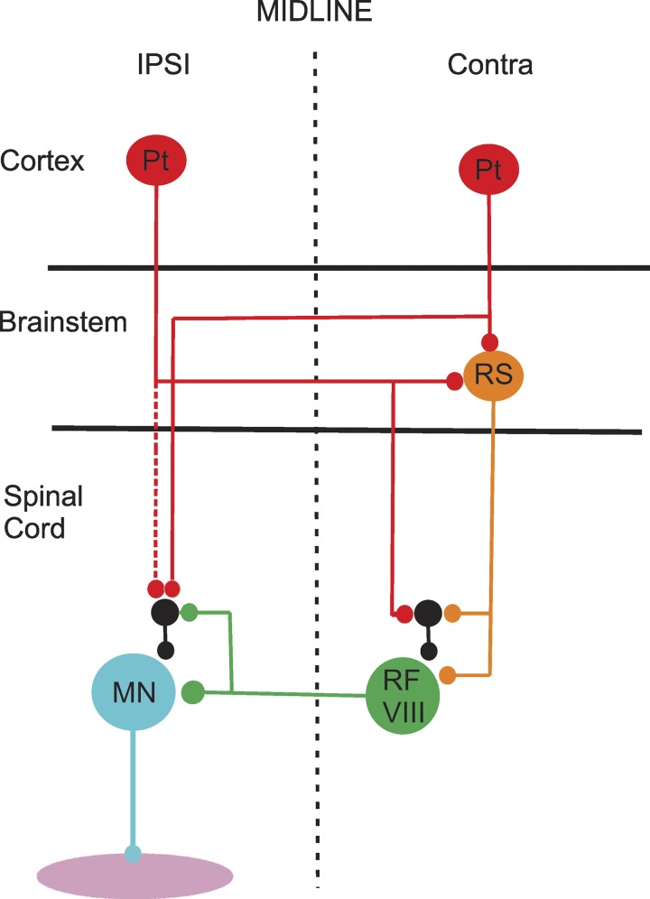
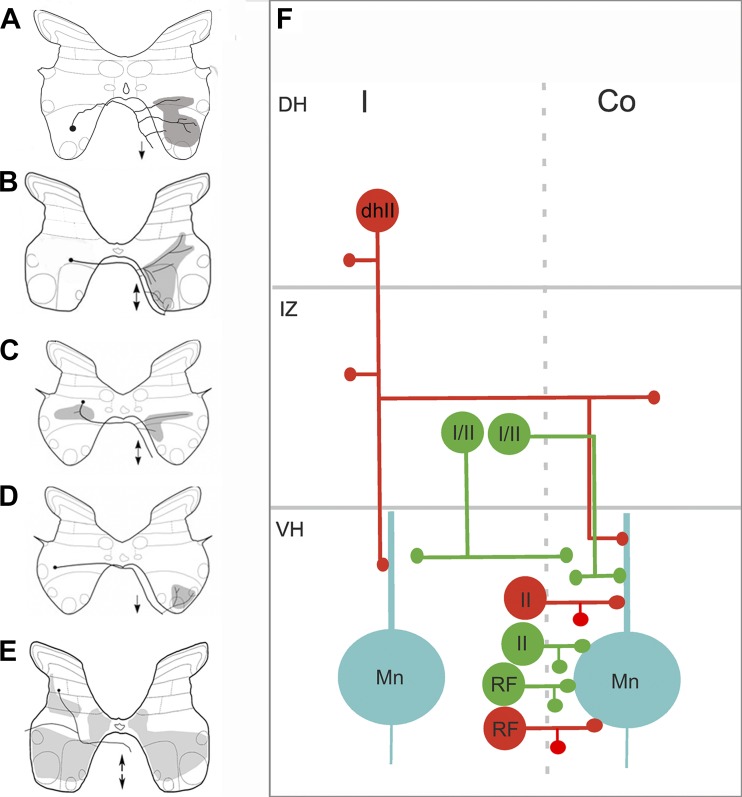
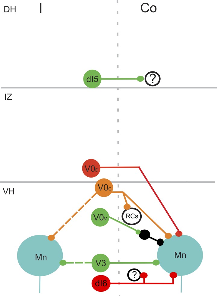
Commissural systems are essential components of motor circuits that coordinate left-right activity of the skeletomuscular system. Commissural systems are found at many levels of the neuraxis including the cortex, brainstem, and spinal cord. In this review we will discuss aspects of the mammalian spinal commissural system. We will focus on commissural interneurons, which project from one side of the cord to the other and form axonal terminations that are confined to the cord itself. Commissural interneurons form heterogeneous populations and influence a variety of spinal circuits. They can be defined according to a variety of criteria including, location in the spinal gray matter, axonal projections and targets, neurotransmitter phenotype, activation properties, and embryological origin. At present, we do not have a comprehensive classification of these cells, but it is clear that cells located within different areas of the gray matter have characteristic properties and make particular contributions to motor circuits. The contribution of commissural interneurons to locomotor function and posture is well established and briefly discussed. However, their role in other goal-orientated behaviors such as grasping, reaching, and bimanual tasks is less clear. This is partly because we only have limited information about the organization and functional properties of commissural interneurons in the cervical spinal cord of primates, including humans. In this review we shall discuss these various issues. First, we will consider the properties of commissural interneurons and subsequently examine what is known about their functions. We then discuss how they may contribute to restoration of function following spinal injury and stroke.

Keywords: bilateral; commissural; movement.


Conflict of interest statement

No conflicts of interest, financial or otherwise, are declared by the authors.

Figures

Fig. 1.
Fig. 1.
Locations of commissural interneuron cell bodies in the lumbar and cervical spinal cord. Commissural interneuron (CIN) cell bodies are shown on the left side of transverse sections and all injections were made on the right side. A: CINs labeled with the b subunit of cholera toxin (CTb) in L5 of the adult rat spinal cord. CTb was injected into the contralateral L3 intermediate gray matter. B: distribution of cells labeled as in A made from eight sections from L5. Adapted from Liu et al. (2010) with permission from Elsevier. Note the clusters of cells in lamina VIII, the medial intermediate gray matter and the lateral deep dorsal horn. C: transverse section of a C4 lumbar segment from a young mouse. Last-order premotor interneurons are labeled transsynaptically with a rabies virus that was injected into the right tibialis anterior muscle. Note the concentration of CINs in lamina VIII (demarcated by yellow dots; A. Bannatyne, B. S. Bhumbra, A. J. Todd, D. J. Maxwell, M. Beato, unpublished observations). D: plot of distribution of Fluorogold-labeled CINs in a single C4 section of an adult rat. Adapted from Mitchell et al. (2016). The black area on the right represents the injection site. E: cells in segment C4 labeled with a retrograde virus [Hiret-GFP (Kinoshita et al. 2012)] that was injected unilaterally into C7–C8 segment of an adult macaque. The area demarcated by the box is shown at higher magnification in F; note the labeled CINs in lamina VIII (A. Bannatyne, E. Bagdatlioglu, D. J. Maxwell, D. S. Soteropoulos, unpublished observations).
Fig. 2.
Fig. 2.
Neurotransmitters associated with commissural interneuron (CIN) axon terminals in rat lumbar motor nuclei. A and A′ show general overviews of CIN terminals and their neurotransmitter content before (A) and after (A′) a sequential reaction with an antibody against choline acetyltransferase to identify motoneurons (additional green structures in A′). Details of the areas demarcated by the boxes are shown in B1B4. B1B4: single optical sections illustrating the neurotransmitter content of terminals that were labeled with the b subunit of cholera toxin (CTb; shown in red). Immunoreactivity for the vesicular GABA transporter (VGAT), which labels GABAergic and glycinergic terminals) is shown in green, and immunoreactivity for vesicular glutamate transporters (VGLUT1/2) is blue. B4 shows a merged image confirming the immunocytochemical characteristics of the terminals examined. Arrows in B1B4 indicate CTb-labeled excitatory terminals that were positive for VGLUT but negative for VGAT. Arrowheads in B1B4 indicate CTb-labeled inhibitory terminals that were positive for VGAT but negative for VGLUT1/2. The larger arrows in B1B4 indicate a CTb-labeled terminal without immunolabeling for either VGAT or VGLUT. Reprinted from Liu et al. (2010) with permission from Elsevier. Scale bars: A = 10 µm; B1B4 = 5 µm.
Fig. 3.
Fig. 3.
Contacts on commissural interneuron (CINs) from descending systems. A: commissural interneuron in lamina VIII (green) of the rat cervical spinal cord. Red structures are terminals of reticulospinal (RS) neurons. The area within the box is shown at a larger magnification in the series of images on the right. A1 shows the dendrite of the cell. A2 shows RF terminals. A3 shows immunoreactivity for the vesicular glutamate transporter 2. A4 is a merged image showing that several glutamatergic RF terminals contact the dendrite (arrows). Adapted from Mitchell et al. (2016). Scale bars A = 20 µm; A1A4 = 5 µm. B: a dendrite of a lamina VIII commissural cell injected with Neurobiotin in the cat lumbar cord (blue). Red structures are serotonin-immunoreactive terminals and green terminals are dopamine-β hydroxylase immunoreactive; a marker for noradrenergic terminals. Several serotonin terminals make contacts with the dendrite (B1 and B2). From Hammar et al. (2004). Scale bars: B = 5 µm; B1 and B2 = 2.5 µm.
Fig. 4.
Fig. 4.
Properties dorsal horn commissural interneurons activated by group II muscle afferents. A shows an intracellular recording from a cell (top traces). An excitatory postsynaptic potential (EPSP) was evoked by stimulation the sartorius nerve at five times the threshold for the most sensitive fibers but no EPSP could be evoked at twice the threshold. The latency (3rd dotted line) is consistent with monosynaptic activation from group II fibers. Bottom traces show the cord dorsum potentials. The calibration pulse = 0.5 mV and 2 ms. B is a reconstruction of a group II activated cell that was intracellularly labeled with Neurobiotin. Dendrites are shown in red and the axonal arbor in black. Note the extent of the axonal projections within the ipsilateral gray matter and the contralateral ventral horn. C shows images of labeled terminals from the cell (red), immunoreactivity for the glycine transporter T2 (green), and a merged image. The terminals are associates with immunoreactivity and hence are glycinergic. D shows a terminal (red), immunoreactivity for gephyrin (green), and immunoreactivity for choline acetyltransferase (blue). The terminal shown in the box opposes gephyrin puncta, which are associated with the postsynaptic densities of inhibitory synapses and a motoneuron (*) in the contralateral motor nucleus. Adapted from Bannatyne et al. (2006); © 2006 Society for Neuroscience.
Fig. 5.
Fig. 5.
Organization of corticospinal and reticulospinal inputs to lamina VIII commissural interneurons (CINs). The diagram shows how both ipsilateral (IPSI) and contralateral (Contra) pyramidal tract (Pt) cells can indirectly influence the activity of lamina VIII commissural interneurons (RF VIII) via the reticulospinal (RS) tract. The commissural cells project directly to motoneurons (MN), which can also be indirectly influenced from Pt cells and RF CINs. Black cells represent interneurons with ipsilateral projections to motoneurons and CINs. Based on data from Stecina et al. (2008a).
Fig. 6.
Fig. 6.
Projections of identified commissural interneurons (CINs) in cat midlumbar segments. AE: cartoons illustrating typical projections of cells [adapted from Bannatyne et al. 2003, (© 2006 Society for Neuroscience); 2009; Jankowska et al. 2009]. The black dot represents the location of the cell body; black lines show axonal projections, and shaded gray areas show areas of the gray matter innervated by axon terminals. Double-headed arrows in B, C, and E indicate axons that bifurcate and ascend and descend in the contralateral ventral funiculus. Single-headed arrows in A and D indicate descending axons. A: lamina VIII cell monosynaptically activated by reticulospinal fibers. B: a group II-activated lamina VIII cell. C: a group I/II cell in the intermediate gray matter with bilateral projections. D: a group I/II cell with a contralateral projection. E: a dorsal horn group II activated cell with extensive ipsilateral and contralateral projections. F: a schematic diagram showing these projections. Inhibitory cells are in red and excitatory cells are in green. Co, contralateral; DH, dorsal horn; dhII, dorsal horn group II activated cell; I, ipsilateral; I/II, cells in the intermediate gray activated by group I and II afferent fibers; II, lamina VIII group II activated cell; IZ, intermediate zone; Mn, motoneuron; RF, lamina VIII cell activated by reticulospinal axons; VH, ventral horn.
Fig. 7.
Fig. 7.
Organization and projections of various classes of commissural interneuron (CIN) according to embryological origin. Glutamatergic dI5 cells settle in the deep dorsal horn (DH) and project to the contralateral dorsal horn. Their target neurons are unknown. V0D cells are inhibitory. They settle within the intermediate gray matter (IZ) or ventral horn (VH) and project to motoneurons (Mn). V0C are cholinergic and found in lamina X and VII close to the midline. Some of them project bilaterally to contralateral and ipsilateral motoneurons and may have additional projections to Renshaw cells (RCs). V0V cells are excitatory and project to motoneurons and Ia inhibitory interneurons (Ia INs). V3 cells are excitatory and have bilateral projections to ipsilateral and contralateral motoneurons. Inhibitory dI6 cells are present in laminae VII and VIII. They project to contralateral motoneurons and additional ventral horn cells. Green, excitatory cells; red, inhibitory cells; orange, cholinergic cells. Co, contralateral; I, ipsilateral. Based on data from Alaynick et al. (2011); Briscoe et al. (1999); Kiehn (2016); Vallstedt and Kullander (2013); Zagoraiou et al. (2009).
Fig. 8.
Fig. 8.
Crossed responses in motoneurons supplying intrinsic hand muscle. A: antidromic activation from ulnar nerve at the wrist. Inset on the right shows location of intraspinal electrode eliciting the crossed response. B: mean intracellular response to contralateral intraspinal microstimulation (100-μA stimulus current). C: single-sweep intracellular potentials that contributed to the average in B. D: averaged cord dorsum potential response recorded simultaneously as B. Arrows indicate time of stimulation. E: same as A, but for a different motoneuron from an intrinsic hand muscle. F: mean intracellular responses to stimulation of the contralateral median nerve at the arm (3× motor threshold). The top trace shows the lack of response to a single shock the middle trace shows the EPSP response to a double shock to the contralateral nerve and the lowest trace shows the response to a triple shock to the contralateral nerve. G: averaged cord dorsum potential response recorded simultaneously as F for triple shock stimulation of the contralateral median nerve. Modified from Soteropoulos et al. (2013).
Fig. 9.
Fig. 9.
Interaction of brainstem descending pathways with commissural circuits in the primate. A: intracellular records of responses of a motoneuron from the lower cervical cord in a primate. The green trace shows the mean response to stimulation of the brainstem descending pathways alone (train of 3 shocks), the red trace shows the response to stimulation of the contralateral spinal cord (cISMS) while the blue trace shows the response to combined brainstem and cISMS stimulation. The black trace underneath shows the net response to the cISMS after brainstem responses have been subtracted. Note the presence of the excitatory postsynaptic potential (EPSP) to cISMS that was absent to cISMS stimulation alone (red). The black triangles indicate the time of brainstem stimulation and the gray arrow shows the time of spinal stimulation. B: same as A but for a different motoneuron (MN). C: schematic showing the stimulated pathways and circuits that could be mediating the conditioned response. CIN, commissural interneuron; RST, reticulospinal tract. Modified from Soteropoulos et al. (2013).

Similar articles

Cited by

References

    1. Aggelopoulos NC, Burton MJ, Clarke RW, Edgley SA. Characterization of a descending system that enables crossed group II inhibitory reflex pathways in the cat spinal cord. J Neurosci 16: 723–729, 1996. doi:10.1523/JNEUROSCI.16-02-00723.1996. - DOI - PMC - PubMed
    1. Alaynick WA, Jessell TM, Pfaff SL. SnapShot: spinal cord development. Cell 146: 178–178.e1, 2011. doi:10.1016/j.cell.2011.06.038. - DOI - PMC - PubMed
    1. Alstermark B, Isa T, Ohki Y, Saito Y. Disynaptic pyramidal excitation in forelimb motoneurons mediated via C3–C4 propriospinal neurons in the Macaca fuscata. J Neurophysiol 82: 3580–3585, 1999. doi:10.1152/jn.1999.82.6.3580. - DOI - PubMed
    1. Alstermark B, Isa T, Pettersson LG, Sasaki S. The C3-C4 propriospinal system in the cat and monkey: a spinal pre-motoneuronal centre for voluntary motor control. Acta Physiol (Oxf) 189: 123–140, 2007. doi:10.1111/j.1748-1716.2006.01655.x. - DOI - PubMed
    1. Alstermark B, Kümmel H. Transneuronal transport of wheat germ agglutinin conjugated horseradish peroxidase into last order spinal interneurones projecting to acromio- and spinodeltoideus motoneurones in the cat. 1. Location of labelled interneurones and influence of synaptic activity on the transneuronal transport. Exp Brain Res 80: 83–95, 1990. doi:10.1007/BF00228850. - DOI - PubMed

Publication types