. 2020 Mar;161(3):595-608.
doi: 10.1097/j.pain.0000000000001745.

Implications of the putamen in pain and motor deficits in complex regional pain syndrome

Affiliations

Implications of the putamen in pain and motor deficits in complex regional pain syndrome

Monica Azqueta-Gavaldon et al. Pain. 2020 Mar.

Abstract

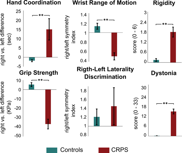
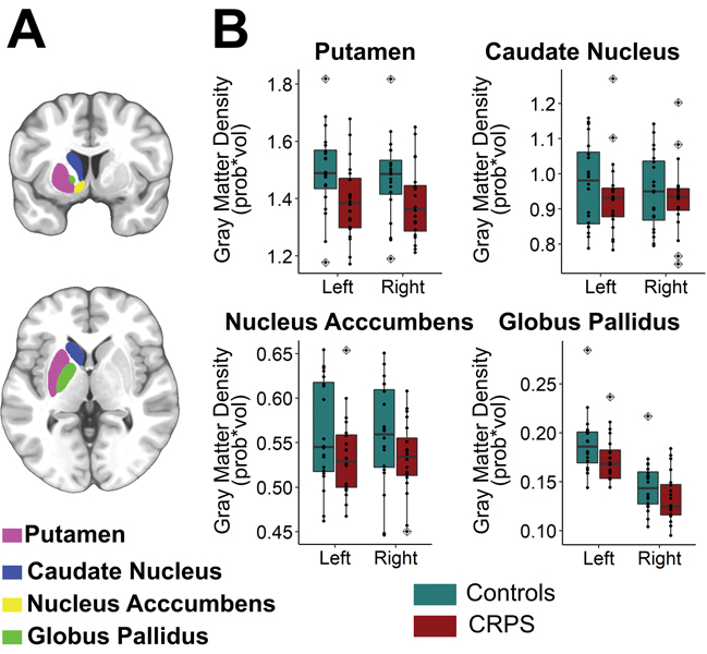
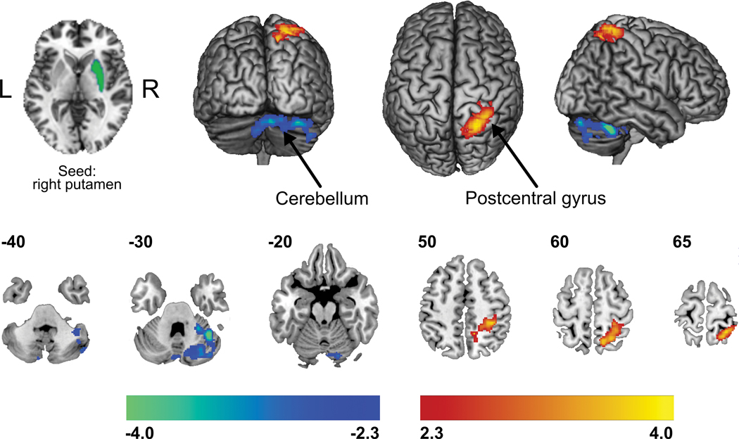
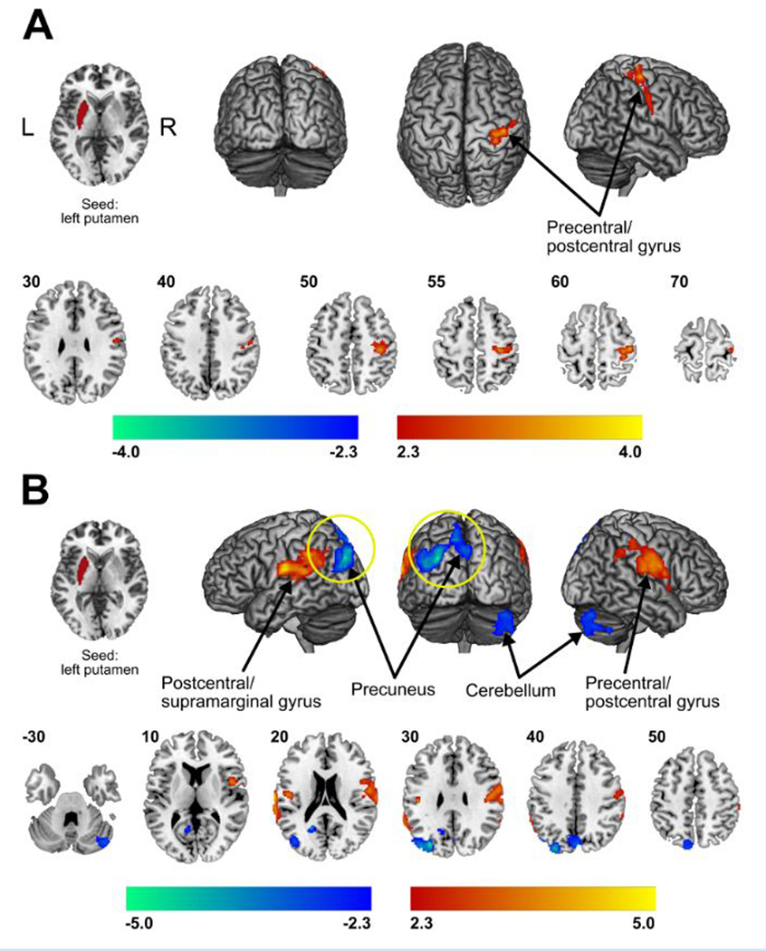
Complex regional pain syndrome (CRPS) develops after-limb injury, with persistent pain and deficits in movement frequently co-occurring. The striatum is critical for mediating multiple mechanisms that are often aberrant in CRPS, which includes sensory and pain processing, motor function, and goal-directed behaviors associated with movement. Yet, much remains unknown with regards to the morphological and functional properties of the striatum and its subregions in this disease. Thus, we investigated 20 patients (15 female, age 58 ± 9 years, right-handed) diagnosed with chronic (6+ months of pain duration) CRPS in the right hand and 20 matched, healthy controls with anatomical and resting-state, functional magnetic resonance imaging. In addition, a comprehensive clinical and behavioral evaluation was performed, where each participant's pain, motor function, and medical history were assessed. Complex regional pain syndrome patients harbored significant abnormalities in hand coordination, dexterity, and strength. These clinical pain- and movement-related findings in CRPS patients were concomitant with bilateral decreases in gray matter density in the putamen as well as functional connectivity increases and decreases among the putamen and pre-/postcentral gyri and cerebellum, respectively. Importantly, higher levels of clinical pain and motor impairment were associated with increased putamen-pre-/postcentral gyri functional connectivity strengths. Collectively, these findings suggest that putaminal alterations, specifically the functional interactions with sensorimotor structures, may underpin clinical pain and motor impairment in chronic CRPS patients.


Conflict of interest statement

The authors declare that there is no conflict of interest regarding the publication of this article.

Figures

Figure 1.
Figure 1.. Functional motor tests performance.
CRPS patients presented with significantly (**p < 0.01) substantial motor deficits in the affected hand relative to healthy controls. The functional motor metrics reflect the performance of the affected (dominant, in case of healthy controls) hand in relation to the healthy (non-dominant) hand to account for intragroup variability.
Figure 2.
Figure 2.. Box plots to show group differences in gray matter density.
Out of all of the striatal ROIs analyzed (A), only the putamen showed a significant bilateral reduction in GMD in chronic CRPS patients when compared to healthy controls. Black dots indicate singe-subjects data. Outlier observations (e.g: outside 1.5 times the interquartile range above the upper quartile and bellow the lower quartile) are denoted with a rhombis symbol. Statistical results stemming from group x side repeated measures ANOVA are given in Table 3.
Figure 3.
Figure 3.. Group differences in resting state functional connectivity of the putamen.
Compared to matched controls, CRPS patients demonstrated greater functional connectivity strength (warm colors) amongst the right (ipsilateral to the affected limb) putamen and sensorimotor and superior parietal cortices, while decreased connectivity (cold colors) was quantified with the crus I region of the cerebellum. Statistical maps were thresholded at p-value < 0.05, corrected for multiple comparisons. Color bars show z-values.
Figure 4.
Figure 4.. Correlation of resting state functional connectivity of the putamen with behavioral measures.
Patients reporting higher spontaneous pain intensity on the day of the scan showed greater functional connectivity of the left putamen with spread motor and sensory discriminative areas. The pain intensity was reported on a visual scale of 0 = “no pain” to 10 = “worse imaginable pain” (A). Interaction analysis (diseaseXmotor dysfunction) showed that patients with poorer hand and finger coordination, as evaluated with the 9-Hole Peg Test, presented increased functional connectivity strengths (warm colors) amongst the left putamen and motor and discriminative/association areas, as well as decreased functional connectivity (cold colors) between the left putamen and both the cerebellum (crus I and II) and precuneus (B). Statistical maps were thresholded at p-value < 0.05. Color bars show z-values. Further Correction for multiple comparison using false discovery rate (FDR) showed that only decreases of functional connectivity of putamen with precuneus in the context of poor hand and finger coordination were significant – highlighted with a yellow circle in the figure.

References

    1. Ab Aziz CB, Ahmad AH. The role of the thalamus in modulating pain. Malaysian J Med Sci 2006;13:11–18. - PMC - PubMed
    1. Alshelh Z, Marciszewski KK, Akhter R, Di Pietro F, Mills EP, Vickers ER, Peck CC, Murray GM, Henderson LA. Disruption of default mode network dynamics in acute and chronic pain states. NeuroImage Clin 2018;17:222–231. doi:10.1016/j.nicl.2017.10.019. - DOI - PMC - PubMed
    1. Andersson JLR, Jenkinson M, Smith S, Andersson J. Non-linear registration aka Spatial normalisation FMRIB Technial Report TR07JA2. 2007. p. Available: http://www.fmrib.ox.ac.uk/datasets/techrep/tr07ja2/tr07ja2.pdf Accessed 14 Nov 2017.
    1. Apkarian AV, Bushnell MC, Treede R-D, Zubieta J-K. Human brain mechanisms of pain perception and regulation in health and disease. Eur J Pain 2005;9:463–84. doi:10.1016/j.ejpain.2004.11.001. - DOI - PubMed
    1. Ashburner J. A fast diffeomorphic image registration algorithm. Neuroimage 2007;38:95–113. doi:10.1016/J.NEUROIMAGE.2007.07.007. - DOI - PubMed

MeSH terms